Dissection 02 – Trunk Musculature, Vertebral Column and Spinal Cord
Ligaments of the Vertebral Column and Intervertebral Discs (Independent Study)
Key concept
The ligaments of the vertebral column support the joints of the vertebral column and limit movement of the vertebral column. The intervertebral discs are composed of the anulus fibrosus and the nucleus pulposus.
Learning objectives
- Map the location of the ligaments of the vertebral column and their function.
- Describe the composition of the intervertebral discs and their function.
Ligaments of the Vertebral Column
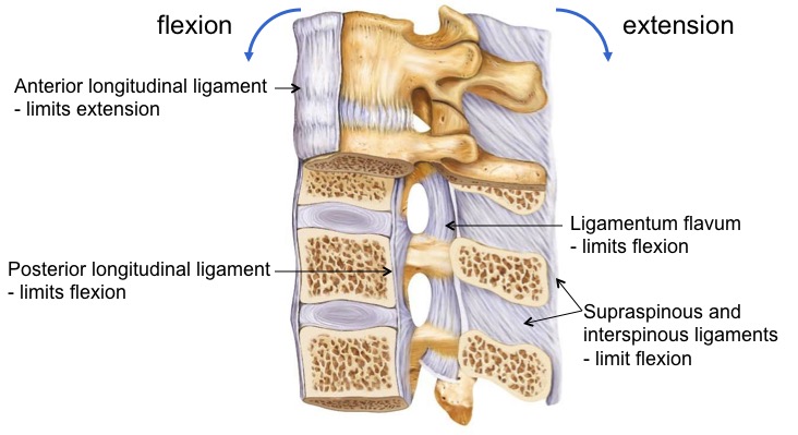
Ligaments work when they become taut. When a ligament is stretched, it is pulled tight and limits whatever movement is pulling it tighter. When thinking about which movement a ligament limits, think about which movement (in this case, flexion or extension) is going to make it taut.
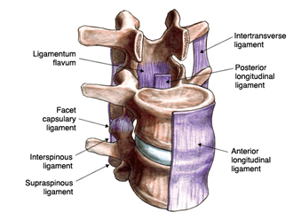
Several ligaments limit the amount of movement at the intervertebral joints. The first two longitudinal ligaments join all of the vertebral bodies.
- Anterior longitudinal ligament: This strong, broad ligament extends along the entire length of the vertebral column covering the anterior 1/3 of each body. It limits extension (prevents hyperextension) and reinforces the anterior portion of the intervertebral disc.
- Posterior longitudinal ligament: It is narrower and weaker than the anterior longitudinal ligament. It lies on the anterior wall of the vertebral canal (posterior surface of the vertebral body). It is narrow as it crosses the posterior surfaces of the vertebral bodies but flares out to blend with the anulus fibrosus of the intervertebral discs. However, it incompletely reinforces the anulus fibrosus posteriorly. Consequently, intervertebral disc herniation is usually posterolateral, where the anulus is not reinforced. This ligament limits flexion.
- Ligamentum flavum: This ligament links the laminae of adjacent vertebrae along the posterior wall of the vertebral canal. It is unique because of the high content of yellow elastic tissue. The ligamentum flavum is stretched, similar to a rubber band, during flexion of the vertebral column and its elastic recoil assists in extension of the column.
- Supra- and interspinous ligaments: These attach spinous processes of adjacent vertebrae and prevent hyperflexion. They are usually very weak in humans. During a midline approach for a spinal tap, the needle traverses these ligaments.
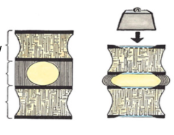
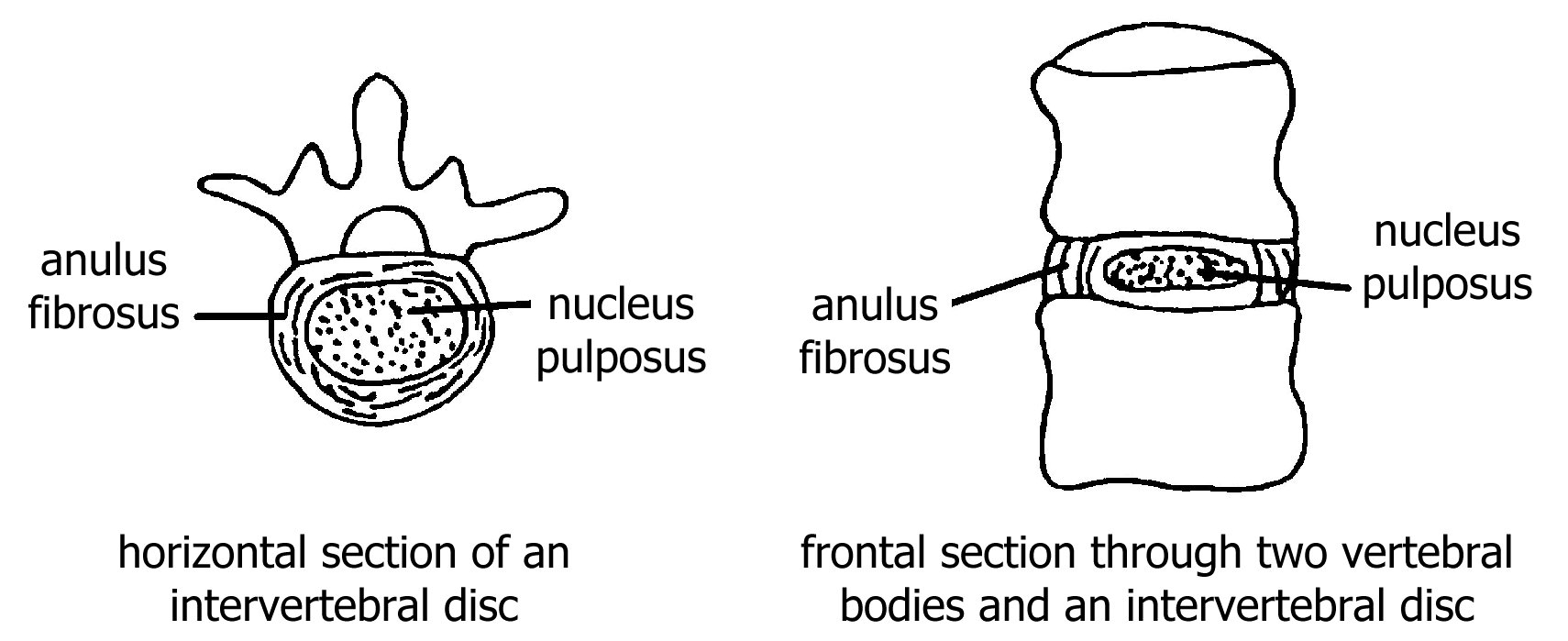
VI. Intervertebral Disc Structure
Intervertebral discs separate the bodies of all adjacent vertebrae with one exception. There is not an intervertebral disc between the atlas and axis. The discs are composed of a peripheral area of fibrocartilage called the anulus fibrosus, and a core of gelatinous tissue called the nucleus pulposus. Both change shape when compressed; the nucleus pulposus serves as a gel-like structure that absorbs forces impacting the vertebrae. The thickness of the disc affects the amount of movement that is possible between vertebrae. The thicker the disc, the greater the movement. Discs are thickest in the cervical and lumbar regions and contribute to the greater degree of movement at these levels.