Dissection 15 – Orbit
Blood Supply to the Orbit – Independent Study
key concepts
There is one artery that provides blood to orbital contents, but multiple venous drainage pathways for blood to leave the orbit.
Learning objectives
- Describe the structures supplied by branches of the ophthalmic artery and be aware of the following branches:
- nasociliary
- lacrimal
- supraorbital
- Describe the three routes of venous drainage from the tissues of the orbit.
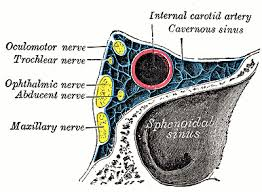
- Identify the five nerves and the artery that pass through the cavernous sinus and what to expect with cavernous sinus syndrome.
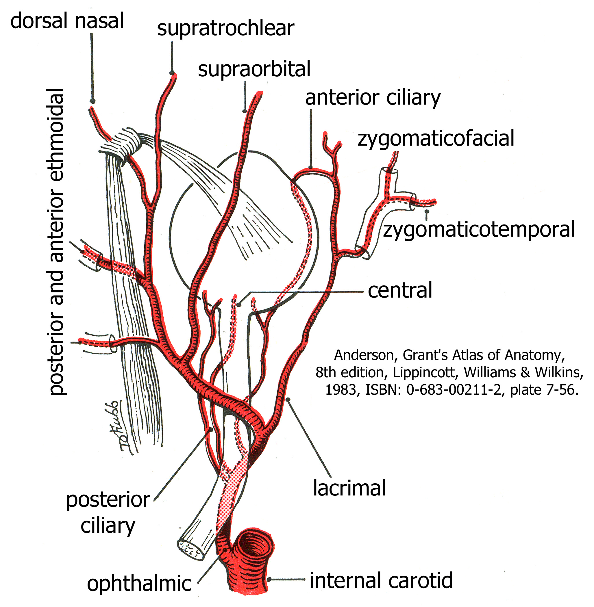
Ophthalmic Artery
The ophthalmic branch of the internal carotid artery supplies tissues in and around the orbit. Although these branches are tiny and very difficult to dissect you should be aware of them. In an atlas, try to identify the lacrimal, supraorbital and nasociliary branches. The ophthalmic artery enters the orbit through the optic foramen. The branches include:
- central artery of the retina through the optic nerve and continues forward into the eye to supply the retina, the inner layer of the eye
- ciliary arteries to the outer layers of the eye
- lacrimal artery to the lacrimal gland
- muscular branches to the extraocular muscles
- ethmoidal arteries to the ethmoidal sinuses and nasal cavity
- the dorsal nasal, supratrochlear and supraorbital arteries to tissues of face and anastomose with branches of the external carotid artery
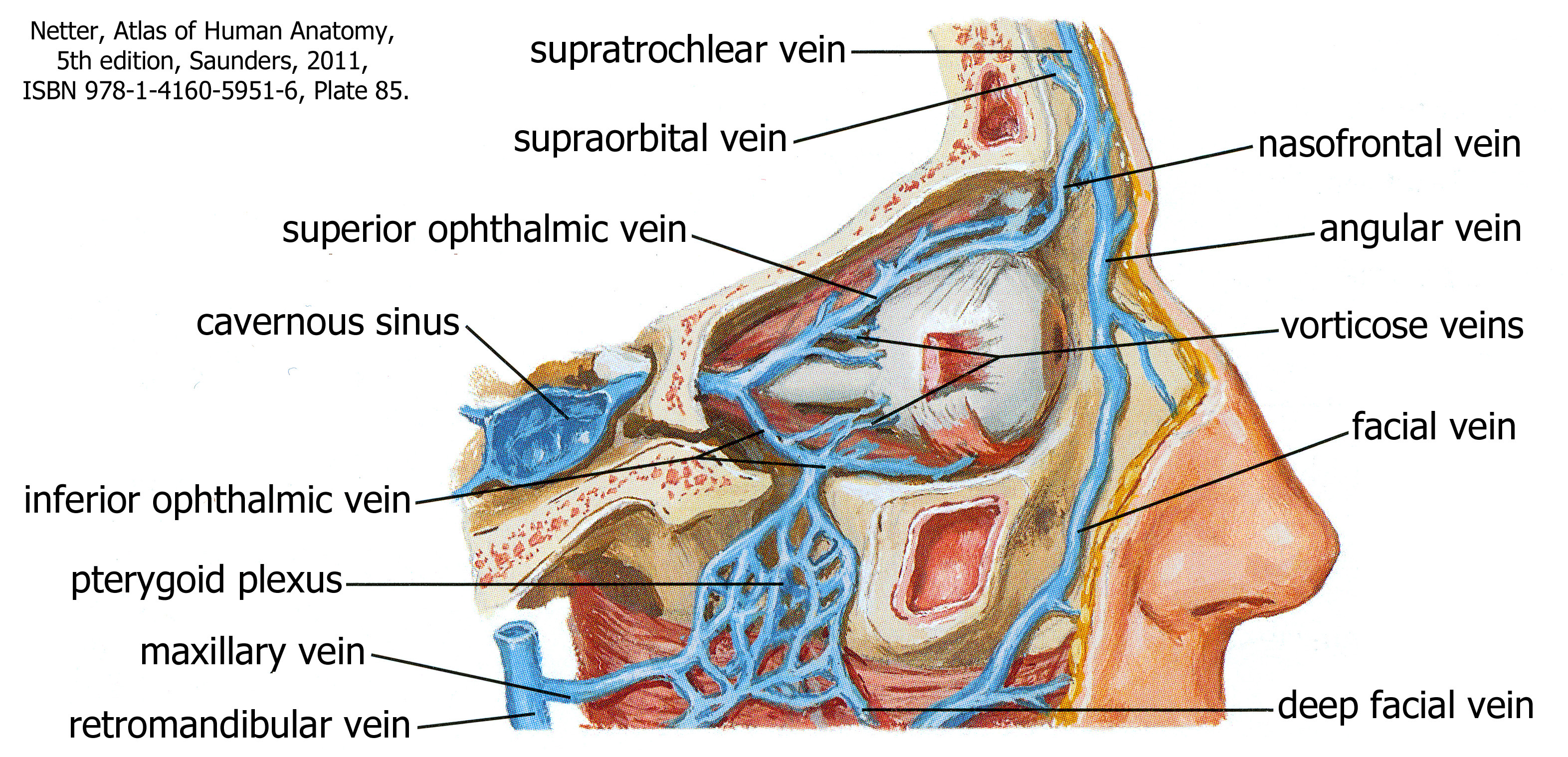
Venous Drainage of the Orbit
The inferior and superior ophthalmic veins usually drain blood from the orbit posteriorly to the cavernous sinus. However, there are two other alternatives. The ophthalmic veins can drain anteriorly to the angular vein of the face, or inferiorly into the pterygoid plexus in the infratemporal fossa. Because of these connections and the fact that these veins are not valved, blood can flow in any direction through these veins, which is important when considering the spread of infection. It is important to note that the cavernous sinus is quite large. Five nerves and one artery travel through the cavernous sinus to enter the orbit in some capacity. Because infection can spread through venous blood from the face (think pimple) into the cavernous sinus and the venous sinuses of the brain, the nerves that travel through the cavernous sinus can, too, be affected by an infection from the face. Presentation of cavernous sinus syndrome is with palsies of whichever cranial nerve is affected (impairment of eye movement, Horner’s syndrome).