
4 Abdominal Quadrants | Digestive System | Abdominal Radiology
Learning Objectives:
By the end of this lab, students will:
- Identify the vasculature of the digestive system and accessory organs including the great vessels and associated branches.
- Identify the structures of the alimentary canal (digestive tract) and the accessory organs of the digestive system.
- Identify the ducts carrying bile from the liver and gallbladder to the duodenum.
- Identify the quadrants of the abdomen and list the organs found in each quadrant.
- Identify structures of the abdominal cavity using various imaging modalities.
Terms to Know
|
Vasculature
Digestive Tract
|
Accessory Digestive Organs
Abdominal Quadrants
|
Introduction
In this lab, you will explore the organs of the digestive system located in the thorax, abdomen, and pelvis. This includes both the organs of the alimentary canal or digestive tract, as well as the accessory digestive organs. Though the spleen is not part of the digestive tract, we will discuss it in this lab due to its location in the abdomen. You will also organize the organs into their abdominal quadrants.
Lab Activities
Activity 1: Abdominal Quadrants Exercise
How do you evaluate the location of internal organs when you can’t see them? It is of critical importance for clinicians in a variety of health science fields to navigate the location of these organs when looking at the abdomen. Where to palpate (feel with your hands), what type of pain is produced, and does pain move or extend into other quadrants are all key questions a clinician may want to know. Fortunately, the umbilicus (aka your belly button) sits right in the center and allows us to create a grid or quadrant. We orient by designating the quadrants by anatomical location (not the way YOU look at them, but how they sit with respect to the patient’s right & left sides): Upper Right Quadrant (URQ), Upper Left Quadrant (ULQ), Lower Left Quadrant (LLQ), and Lower Right Quadrant (LRQ). The umbilicus (belly button) represents where each line crosses.
Activity 2: Digestive System Organs
Watch the videos on Canvas that explore the organs of the digestive system as well as the spleen. There are several videos, with some showing the cadaveric tissue and others using the digital atlas. Be sure to watch all of the videos, as some structures are not visible using one modality or another (tissue vs. atlas). Observe the structures of the digestive system in the images provided in the Abrahams‘ and Netter’s atlases as well.
- Observe the spleen. This structure is part of the lymphatic system, not the digestive system, but we will examine it today due to its location in the superior abdomen, surrounded by digestive organs. This organ recycles old blood cells, kills bacteria and other foreign particles, and plays an important role in the immune system.
- Observe the sections of the liver. The liver is involved in many functions, including bile production; detoxification of drugs, metabolites, and poisons; storage of certain vitamins and nutrients, and synthesis of blood plasma proteins.
- Identify the lobes of the liver. There is usually a thin layer of tissue that separates the right and left lobes. On the inferior aspect, you can identify the caudate (posterior) and quadrate (anterior) lobes. The vasculature is located on the posterior and inferior aspects of the liver. The largest, thin-walled vessel is the inferior vena cava. This vessel ascends through the abdomen and carries blood from the lower extremities and trunk back to the heart. It picks up blood that has been processed in the liver along its path. Normally on the inferior surface, you will see three vessels near each other. The smaller but thicker-walled vessel is the common hepatic artery. This artery supplies oxygenated blood to the tissue of the liver. This is a branch of the celiac trunk, which you will learn more about in the next lab. In the same region, you will see a larger, thinner-walled vessel. This is the hepatic portal vein. This vein carries blood from the digestive tract to the liver so that it can be processed and detoxified before entering general circulation. The final vessel you will see in this region is the common hepatic duct, which carries bile from the liver to the duodenum.
- Observe the regions of the large intestines: the cecum, ascending colon, transverse colon, descending colon, and sigmoid colon. The primary function of the large intestines is the absorption of water. The small tube-like structure extending from the cecum is the appendix. Though its function was misunderstood for a long time, we now know that the appendix has lymphatic and immune functions.
- Observe the esophagus, stomach, and proximal duodenum. The esophagus is located along the midline, just to the right of the descending aorta, posterior to the trachea (superiorly) and just anterior to the vertebral column. The esophagus has to pass through the posterior aspect of the diaphragm in order to reach the stomach. There is a subtle thickening where the esophagus meets the stomach. This is the inferior esophageal sphincter, and it works to prevent regurgitation of stomach contents back into the esophagus.
- Now observe the stomach. In the video and some images it has been opened and the rugae, or folds on the interior surface, are clearly visible. Identify the different regions of the stomach. The cardiac region is nearest the esophagus, and the fundus is the most superior, dome-shaped region. Though the borders are difficult to ascertain, the central region of the stomach can be considered the body. Near the entrance to the duodenum is the pyloric region of the stomach. Where the stomach approaches the duodenum, there is a very thick sphincter called the pyloric sphincter. This regulates the passage of digested materials from the stomach to the small intestines.
- Observe the artery that is twisting and winding near the pancreas. This is the splenic artery. It is a branch of the celiac trunk, and it supplies the spleen, part of the stomach, and part of the pancreas. The second branch of the celiac trunk is the left gastric artery, and it supplies the rest of the stomach and inferior esophagus.
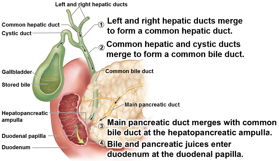
- Observe the gallbladder, which is typically green. The gallbladder stores bile that has been produced by the liver. Bile is carried from the liver through the common hepatic duct. The cystic duct connects the common hepatic duct to the gallbladder. The duct formed by the merging of the common hepatic duct and cystic duct is the common bile duct. When food enters the small intestines, bile travels from the liver via the common hepatic duct and gall bladder through the cystic duct, then through the common bile duct to the duodenum. Bile plays an important role in fat digestion.
- Observe the common bile duct, common hepatic artery, and hepatic portal vein traveling together towards the liver would be (near the gallbladder). The common hepatic artery is the third branch of the celiac trunk, and it supplies the liver as well as parts of the stomach and duodenum. While this artery is supplying the liver with oxygenated blood, the hepatic portal vein is supplying it with blood from the digestive system containing nutrients absorbed there.
- Observe the pancreas. It is light yellow in color and more granular than nearby adipose (fat) tissue. Pancreatic endocrine cells secrete insulin and glucagon into the blood to regulate blood sugar levels. Pancreatic exocrine cells produce enzymes and bicarbonate that are key for digestion in the small intestines. The pancreatic duct joins the common bile duct at the hepatopancreatic ampulla just before both empty their contents into the duodenum. Follow the common bile duct to the duodenum. From there, you can follow the pancreatic duct a short ways to the pancreas. Notice how the hepatopancreatic ampulla, where these ducts meet, is very short and somewhat enlarged. The duodenal papilla, sometimes visible on the internal aspect of the duodenum, is where these ducts empty their contents into the duodenum.
- Also observe the greater omentum, which lies over the anterior aspect of the abdominal organs. The greater omentum is one of the mesenteries of the abdominal cavity. The mesenteries hold organs in place, store fat, and provide a route for vessels and nerves to reach the organs.
Activity 3: Cross-sectional anatomy of the digestive system
Watch the video on Canvas that reviews the cross-sectional anatomy of the abdominal cavity. In living patients, we often view organs of the abdominal cavity in cross-section via CT or MRI images. If you can understand the cross-sectional anatomy in the color images in the video, it will be easier to understand what you are seeing in the radiology images.
Activity 4: Abdominal Cavity Radiology (Digestive Organs and spleen)
View the radiology video in Canvas presenting on the radiology of the abdominal cavity, specifically the digestive organs and spleen. The slides will also be posted as a study tool. Observe the following structures in the radiology images.
- Stomach
- Pancreas
- Liver
- Small intestines
- Ascending colon
- Transverse colon
- Descending colon
- Hepatic flexure
- Splenic flexure
- Spleen
- Abdominal Aorta
- Inferior vena cava
wRAP UP AND Clinical Connection
For today’s clinical connection, we will review the imaging of a liver laceration and cirrhosis of the liver.
