
8 Muscles and Triangle of the Neck and Face | Vasculature of the Head and Neck
Learning Objectives:
- Identify the muscles of the neck and face, their actions, and their innervation.
- Describe the vasculature of the head and neck and identify the regions supplied or drained by each vessel, as described in the lab guide.
- Review the cross-sectional anatomy of the brain through radiology images.
Terms to Know
|
The Neck
Arteries of the Head and Neck
|
Veins of the Head and Neck
Muscles of Facial Expression
|
Introduction
In this lab you will explore the muscles and vasculature of the neck and face. These muscles allow us to express emotions from happiness to sadness, surprise to anger. The structures discussed in this lab have a range of clinical implications, from the scalene muscles placing pressure on neurovascular structures to vascular compromise that can be fatal.
Lab Activities
Activity 1: Muscles of FACIAL EXPRESSION
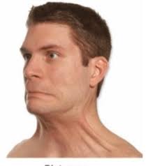
The muscles of facial expression generally originate on the bones and fascia of the skull and insert into the skin of the face, resulting in movement and wrinkling of the facial skin. All of these muscles, except for one (levator palpebrae superioris, CN III), are innervated by the facial nerve, CN VII. A chart is provided in this lab guide to provide you with more information about these muscles. You are responsible for knowing actions and emotions or common motions conveyed by these muscles. You are encouraged to study the description of the muscle actions while viewing the muscles.
While won’t be asked about origins and insertions for these muscles, knowing where they insert and the direction that their fibers run can help you understand their action. For example, two muscles are located superior to the lip, with nearby origins inferior to the eye. Though they both insert into the upper lip and elevate parts of the upper lip, they have different insertion points. As a result of these different insertions, the contraction of these muscles conveys very different emotions. Levator anguli oris inserts into the corner of the mouth, while levator labii superioris inserts medial to that, in the upper lip. Therefore, when levator anguli oris contracts, it causes smiling, while contraction of levator labii superioris conveys sadness by elevating the upper lip. If you are taking the exam and can’t remember one of these muscles’ action, look at the insertion point, which will help you reason through the action.
Observe the muscles of the face:
|
Muscle |
Description |
Action |
Emotion/ Common Motion |
|
|
Occipito-frontalis |
Frontal belly |
Frontal belly covers forehead; occipital belly covers posterior skull; Epicranial aponeurosis connects the bellies |
Elevates eyebrows & wrinkles forehead horizontally |
Surprise, Curiosity |
|
Occipital belly |
Pulls the scalp posteriorly |
|||
|
Corrugator supercilii |
Small muscle in the medial eyebrow |
Draw eyebrows inferomedially, creating vertical wrinkles above the nose |
Concern, skepticism |
|
|
Orbicularis oculi |
Thin, flat, sphincter of the eye, surrounds the orbit |
Closes eyes |
Winking, blinking; squinting |
|
|
Levator palpebrae superioris*** |
Runs from the posterior orbit to the superior eyelid |
Elevates superior eyelid to open the eye |
Contributes to surprise, curiosity |
|
|
Procerus |
Between the eyebrows over the nasal bone |
Depresses medial eyebrow, wrinkles skin over the nose |
Dislike or disdain |
|
|
Nasalis |
Over cartilage of the nose |
Compresses bridge and depresses tip of the nose, elevates corners of the nostrils |
Flares nostrils, as with anger |
|
|
Buccinator |
Thin, horizontal muscle in the cheek, deep to the masseter. |
Compresses cheek; holds food between cheek and teeth when chewing |
Whistling, sucking |
|
|
Depressor anguli oris |
Runs from the angle of the mouth to the lateral chin |
Draws the corner of the mouth inferiorly and laterally |
Frown |
|
|
Depressor labii inferioris |
Runs from lateral chin to the lower lip |
Depresses the lower lip |
Frown |
|
|
Levator anguli oris |
Runs from the medial cheek to the angle of the lips |
Widens the mouth, elevates the corners of the mouth |
Smiling |
|
|
Levator labii superioris |
Runs from the inferior orbit to the lateral upper lip |
Elevates and furrows the upper lip |
Sadness; |
|
|
Levator labii superioris alequae nasi^ |
Runs alongside nose to medial upper lip, lateral to midline |
Elevates the upper lip, dilates the nostrils |
“Elvis” snarl |
|
|
Zygomaticus major |
Zygomatic arch to the corner of the mouth |
Raises the corners of the mouth |
Smile |
|
|
Zygomaticus minor |
Zygomatic arch to the lateral upper lip |
Raises the corner of the mouth/lateral upper lip |
Smile |
|
|
Orbicularis oris |
Thin muscle surrounding the entrance to the oral cavity |
Closes the lips/mouth; purses and protrudes the lips |
Kissing, whistling |
|
|
Risorius |
Runs horizontally and laterally from the angle of the mouth |
Draws corner of lip laterally and down, tenses the lips |
Frustration, sadness |
|
|
Mentalis |
Anterior chin to the lower lip |
Elevates & wrinkles skin of the chin, protrudes the lower lip |
Pouting |
|
|
Platysma |
Thin muscle within skin of the neck that runs from the clavicular region to the lower mandible and mouth |
Tenses the skin of the inferior face and neck, depresses the jaw |
|
|
**Note: All of these muscles are innervated by the facial nerve (CN VII) except for levator palpebrae superioris, which is innervated by the oculomotor nerve (CN III).
^Fun Fact!: This is the longest name of a muscle in the body.
Activity 2: Muscles of the Neck
Observe the muscles of the neck:
- Just deep to the platysma, you can observe the sternocleidomastoid muscle. This muscle runs from the sternum and clavicle to the mastoid process of the temporal bone. It is primarily innervated by CN XI, the accessory nerve. When only one side contracts, it causes rotation of the head to the opposite side of the muscle that is contracting. Try palpating your own sternocleidomastoid muscles and rotate your head from side to side. The more you rotate your head to the opposite side, the more this muscle becomes prominent in the anterolateral portion of your neck. Bilateral contraction contributes to the flexion of the neck.
- Deep to the trapezius muscle, observe the splenius capitis. Bilateral contraction of this muscle causes extension of the neck. Unilateral contraction (a contraction of this muscle on only one side) can cause either lateral flexion of the neck to the same side or rotation of the neck to the same side.
- Observe the anterior, middle, and posterior scalene muscles. The scalene muscles are deep to the sternocleidomastoid muscle. The scalene muscles run from the cervical vertebrae to the first and second ribs. Their primary action occurs with breathing. They act to elevate the ribs during forced inspiration. Unilateral contraction of these muscles also contributes to lateral flexion of the neck to the same side and rotation of the neck to the opposite side.
Observe the hyoid bone. This bone is inferior to the mandible within the neck. The hyoid is suspended within the neck by muscles and provides an important site of attachment for them.
Infrahyoid muscles
Observe the infrahyoid muscles. As the name implies, these muscles are located inferior to the hyoid. The names of these muscles indicate the attachment points for these muscles. Together these muscles anchor the hyoid and depress both the hyoid and larynx during swallowing and speaking.
- Observe the omohyoid. This muscle is digastric, meaning that it has two muscle bellies. Observe the short, intermediate tendon between the superior and inferior muscle bellies. This tendon is held to the clavicle by a fascial sling. The word part “omo” means scapula, and this muscle runs from the scapula to the hyoid bone.
- View the sternohyoid. This muscle runs from the sternum to the hyoid.
- Observe the sternothyroid and thyrohyoid, which run from the sternum to the thyroid cartilage and thyroid cartilage to the hyoid bone, respectively.
Suprahyoid muscles
Observe the suprahyoid muscles. These muscles are superior to the hyoid, and all have an attachment site on the hyoid. As a group, these muscles form much of the floor of the mouth and elevate the hyoid and larynx in swallowing and speaking.
- Observe the digastric muscle. Note, don’t confuse this with the omohyoid. That is also a digastric muscle, with two muscle bellies. However, this muscle’s name is the digastric muscle. The anterior belly of the digastric runs from the mandible to the hyoid bone and the posterior belly of the digastric runs from the hyoid bone to the temporal bone (near the mastoid process).
- Superior to the posterior belly of the digastric, observe the stylohyoid. This muscle travels from the styloid process of the temporal bone to the hyoid.
- The mylohyoid can be seen just inferior and medial to the mandible. This muscle runs from the mandible to the hyoid and forms the anterior floor of the mouth.
- The geniohyoid runs from the mandible to the hyoid bone, but it is smaller and sits deep to the mylohyoid.
Activity 3: Vasculature of the Face and Neck
Observe the arteries of the neck in the videos on Canvas:
- Observe how the brachiocephalic trunk divides into the subclavian and common carotid arteries. On the left side of the body, these two arteries branch directly off the aortic arch.
- Follow the subclavian artery laterally for a short span and observe the vertebral artery. This artery travels superiorly through the transverse foramen of the cervical vertebrae to supply the brain. You have already viewed this artery as it comes together to form the basilar artery when you studied the circle of Willis.
- Now observe the common carotid artery. This artery travels superiorly through the neck and branches into the internal and external carotid arteries.
- The internal carotid artery continues superiorly without branching in the neck, travels through the skull, and provides the majority of the blood supply to the brain.
- The external carotid artery supplies blood to the scalp, the face, and some structures of the neck. Six arteries branch from the external carotid artery before it terminates by dividing into two arteries. Observe the following arteries as they branch from the external carotid arteries, in order from inferior to superior:
-
-
- Ascending pharyngeal artery: you will not be asked to identify it on the exam. This artery ascends to supply the pharynx, middle ear, and cranial meninges.
- Superior thyroid artery: Runs inferiorly to supply the thyroid gland and larynx.
- Lingual artery: Observe how this artery runs medially to supply the tongue.
- Facial artery: This artery branches just superior to the lingual artery. In fact, sometimes, the facial and lingual arteries branch from the external carotid artery as a common trunk before dividing. It travels along the inferior aspect of the mandible and then wraps around the middle portion of the mandible and ascends through the face. It supplies the submandibular gland, lips, and muscles of the face.
- Angular artery: This terminal branch of the facial artery runs along the nose to the medial corner of the eye.
- Occipital artery: This artery arises from the posterior aspect of the external carotid artery and travels superiorly and posteriorly to supply the posterior scalp and neck.
- Posterior auricular artery: This artery arises from the posterior aspect of the external carotid artery, just superior to the occipital artery. It ascends just anterior to the mastoid process of the temporal bone and posterior to the ear, and it supplies muscles of this region, the parotid gland, and part of the scalp.
- The external carotid artery terminates as it divides into the superficial temporal artery and maxillary artery.
- Superficial temporal artery: This artery travels superiorly to supply part of the scalp and superolateral face.
- Maxillary artery: This is the primary artery supplying the deep structures of the face. It travels anteriorly and medially and gives branches that supply parts of the external ear, the cranial cavity, the scalp, the mouth (including the cheek, teeth, upper and lower jaw, palate, and gums), and the inferior portion of the orbit.
-
Observe the veins of the head and neck:
- View the external jugular vein, which empties into the subclavian vein. This vein drains part of the lateral aspect of the head.
- The internal jugular vein ultimately drains the majority of the blood from the head and neck. The internal jugular vein joins with the subclavian vein to form the brachiocephalic vein. Unlike the arteries of the same name, this happens on both the right and left sides. They both need to carry their blood toward the midline to reach the superior vena cava. However, for the arteries on the left side, the arch of the aorta already travels towards the left, positioning the subclavian and common carotid arteries in the appropriate place where they would normally branch.
Wrap up and clinical connection
The clinical connection for this lab reviews the muscles of facial expression using face yoga. Face yoga is a fantastic way to exercise your muscles of facial expression and release some tension. Please exercise along if you would like, you might realize your muscles are sore the following day from these movements. Doing these exercises in front of a mirror is priceless.
