
6 The Brain
Learning Objectives:
By the end of this lab, students will:
- Explain the directional terms associated with the orientation of the brain in the cranial cavity.
- Describe the layers of tissue that cover the brain and explain their function.
- Describe the structures of the cerebral hemispheres, including the lobs of the brain and their components, using the whole-brain and midsagittally sectioned specimens.
- Explain the various functions associated with identified regions of the brain.
- Describe the Circle of Willis.
Terms to Know
|
Directional Terms
Cerebrum
|
Meninges
Ventricles
Other Structures
Blood Supply to the Brain
|
Introduction
In this lab, you will learn about the brain. The brain is a mass of neurons and glial cells controlling everything from our ability to breathe, speak, and move our limbs, to our personalities, emotions, and higher-level thinking.
It is important to address two additional directional terms that are used with respect to the brain. A change in the long axis of the nervous system occurs between the cerebrum and the brainstem. The long axis of the cerebrum is relatively horizontal, while the long axis of the brainstem and spinal cord is relatively vertical.
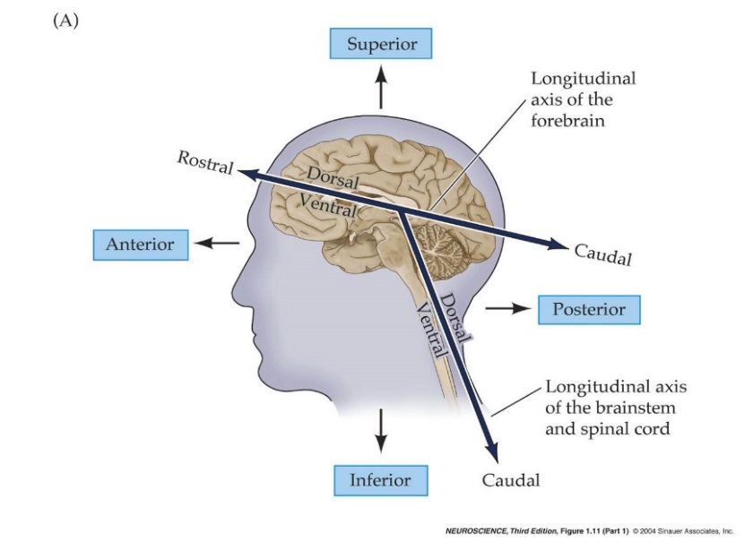
As a result, some of the directional terms may have slightly different meanings depending on the structure to which they refer. Rostral is a term referring to the most frontal portion of the brain (rostrum = nose), while caudal describes structures closer to the tip of the spinal cord (caudal = tail). When used in the context of the cerebrum, rostral refers to structures closer to the nose (as before), but caudal now refers to structures toward the back of the head (posterior). The terms ventral and dorsal can be used interchangeably with anterior and posterior with respect to the plan of the brainstem and spinal cord. However, in the plane of the cerebrum, these terms are interchangeable with inferior and superior.
The University of British Columbia has excellent resources that you may find helpful in studying the brain. These resources at neuroanatomy.ca. The site contains videos, interactive modules, 3D models, cross-sectional images, radiology, and other tools.
Lab Activities
Activity 1: The Meninges
Three connective tissue membranes cover the brain: dura mater, arachnoid mater, and pia mater. The brain has a consistency like somewhat firm jelly during life. The meninges function to protect this soft structure by anchoring it to the skull and preventing excessive movement within the skull.
The dura mater is the thick outer layer of meninges. The dura mater follows the contours of the skull’s inner surface and does not dive into the sulci of the brain. In a few places, it folds and dives into spaces between parts of the brain. These are called the dural reflections. You can explore the dural reflections using the images below.
The arachnoid mater is the middle layer of the meninges, and it is a delicate, transparent membrane that can be seen covering the brain. The arachnoid mater does not follow the contours of the sulci and gyri; rather, it follows the form of the overlying dura. Notice aggregations of tiny white granules near the superior midline. These are arachnoid granulations. They function to return cerebrospinal fluid from the subarachnoid space (between this arachnoid mater layer and the pia mater layer of meninges) to the blood.
The pia mater is the innermost dural membrane. It cannot be seen with a gross examination of the brain, but it covers the surface of the brain tissue, including within the sulci and gyri.
Activity 2: Cerebral Hemispheres and a brief introduction to the cerebellum and brainstem
The cerebral hemispheres contain the brain regions involved in our higher cognitive functions, including language, learning, memory, and personality. Each hemisphere has four lobes: the frontal lobe, temporal lobe, parietal lobe, and occipital lobe. The surface of the cerebral hemispheres is made up of the cerebral cortex, which is a layer of gray matter. This surface is thrown into many folds forming sulci and gyri. The sulci are the folds diving in away from the brain’s visible surface, while the gyri are the portion of the cortex that is exposed and visible. Sulci and gyri are important because they increase the surface area of the cortex, giving us more room for neurons involved in higher cognitive functions.
Explore the following structures of the cerebral hemispheres.
- The median longitudinal fissure separates the left and right hemispheres.
- The corpus callosum is the large white matter pathway connecting the right and left hemispheres of the brain. Observe this structure in cross-section on a midsagittal view or a medial view of a hemisphere.
- The central sulcus marks the boundary between the frontal and parietal lobes. It can be tricky to identify, so to reliably
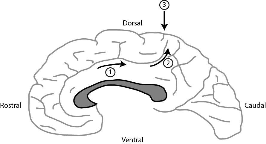 identify it, start from the midsagittal section. Above the corpus callosum (gray in the image to the right), there is a curved gyrus called the cingulate gyrus. The sulcus dorsal to the gyrus is the cingulate sulcus. If you follow the cingulate sulcus (1) in the direction of the arrows, it bends dorsally (2) and comes to a stop very close to the dorsal aspect of the brain surface. Move rostrally one sulcus, and you’ve located the central sulcus (3). Now you can trace it over the lateral aspect of the brain.
 - The precentral gyrus is in the frontal lobe, just anterior to the central sulcus. This is where the primary motor cortex is located, where all voluntary motor signals begin.
- The postcentral gyrus is in the parietal lobe just posterior to the central sulcus. This is the primary somatosensorycortex, which processes general sensory information, including the sense of touch.
- The lateral (Sylvian) fissure is the boundary between the frontal & temporal lobes.
- The temporal lobe contains three gyri running parallel to the lateral (Sylvian) fissure: the superior temporal gyrus, middle temporal gyrus, and inferior temporal gyrus. The primary auditory cortex is located in the superior temporal gyrus.
- The parieto-occipital sulcus separates the parietal lobe from the occipital lobe. It is visible in the midsagittal section.
- The calcarine sulcus (fissure) is in the occipital lobe. It runs roughly perpendicular to the parieto-occipital sulcus and contains the primary visual cortex. It is visible on the midsagittal view.
- The thalamus is located deep in the brain. It can be seen in the midsagittal section in the area of the third ventricle. Many pathways between the cerebrum, brainstem, cerebellum, and spinal cord have a synapse in the thalamus.
- The hypothalamus is located just anterior and inferior to the thalamus. It can also be seen in the midsagittal view. This structure is responsible for many functions, including endocrine control, species-preserving behaviors (hunger, thirst), and circadian rhythm.
- The anterior commissure is located just anterior to the thalamus. It connects parts of the frontal and temporal lobes of the two hemispheres.
Observe the cerebellum and the brainstem’s three parts: the midbrain, pons, and medulla oblongata. You will only see the midbrain on the midsagittal view, as it is mostly hidden by other structures in the whole brain view. For now, be familiar with the general location of these structures. We will discuss them more in-depth in the next lab.
Activity 3: Blood Supply to the Brain – the Circle of Willis
The brain is supplied by two pairs of arteries: the vertebral arteries and the internal carotid arteries. They branch into the arteries that eventually supply the whole brain. Explore these arteries and their branches on the inferior view of the brain.
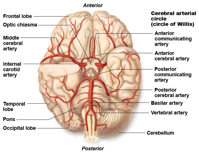
- The vertebral arteries merge along the brainstem to form the single basilar artery.
- The basilar artery travels along the ventral surface of the pons (part of the brainstem). It gives off several branches to the cerebellum along its path.
- The basilar artery splits on the pons’ superior ventral surface to give off right and left posterior cerebral arteries.
- Locate the internal carotid arteries. They travel through the skull and have been cut here, so it looks as if their lumen is facing somewhat towards you.
- The internal carotid arteries divide into two branches: the anterior cerebral artery and the middle cerebral artery. The middle cerebral artery dives into the lateral fissure, while the anterior cerebral artery travels anteromedially to the median longitudinal fissure. Examine a midsagittal section to follow the anterior cerebral artery as it runs along the anterior and superior border of the corpus callosum.
- The posterior communicating arteries are small-diameter arteries that connect the posterior cerebral and internal carotid arteries.
- The two anterior cerebral arteries are connected just before entering the median longitudinal fissure by the anterior communicating artery. This is usually a very short artery connecting them, but sometimes the anterior cerebral arteries can appear to “touch” each other and then split again.
Together the three pairs of cerebral arteries and the three communicating arteries form the Circle of Willis, an arterial circle that provides collateral circulation. There is a great deal of variation in the sizes of the component arteries of the circle, including instances of left-right asymmetry. It is not unusual to have an incomplete circle of Willis.
Activity 4: the ventricles and Cerebrospinal Fluid
The brain contains several ventricles filled with cerebrospinal fluid. Observe the open space in the middle of each hemisphere. These are the lateral ventricles. The septum pellucidum is a thin membrane that separates the anterior part of the lateral ventricles from each other. The larger anterior portion is called the anterior horn of the lateral ventricle. The body of the lateral ventricle is the thinner portion, just posterior to the anterior horn. The portion in the temporal lobe is called the inferior horn of the lateral ventricle. The posterior horn of the lateral ventricle extends posteriorly.
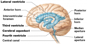
The third ventricle is a thin midline space that separates the left and right thalami. Notice that the third ventricle in the image to the right appears to have a hole in the middle of it. This is created by a midline thalamic structure called the massa intermedia, or interthalamic adhesion, which connects the two thalami and passes through the third ventricle. Each lateral ventricle connects to the third ventricle by way of the interventricular foramen (of Monro).
The third ventricle connects to the fourth ventricle via the cerebral aqueduct. The fourth ventricle lies between the pons and the cerebellum.
All of the ventricles contain choroid plexus, which produces cerebrospinal fluid (CSF) within the ventricles. You may see the choroid plexus in some images, which looks like reddish granular tissue inside the ventricle. CSF can be produced by the choroid plexus in all of the cerebral ventricles, but the longest possible pathway of CSF flow begins in the lateral ventricle. View a midsagittal section and follow the flow of CSF from the production in the choroid plexus in the lateral ventricle through the CNS.
- Lateral ventricle–>Interventricular foramen (of Monro)–>Third ventricle–>Cerebral aqueduct–>Fourth ventricle (From the fourth ventricle, CSF can escape via the lateral apertures, medial aperture, or central canal. You will not be responsible for identifying those structures.)
Activity 5: Cross-sectional Anatomy of the Brain
Observe the following structures in a coronal cross-section view. I recommend using the University of British Columbia Functional Anatomy Cross Sections tool and the cross-section images posted on Canvas to study. The tissue in the cross-section images on Canvas are prepared using a special stain that makes the white matter appear very dark, and the grey matter appears lighter. The UBC Functional Anatomy images show the white and grey matter with its typical coloration. A list of the structures that can be seen on the cross-sections can also be found on the Canvas page.
- *Median longitudinal fissure: Space between the two hemispheres superiorly.
- Corpus callosum: Can be observed as the thick structure connecting the two hemispheres just inferior to the median longitudinal fissure.
- **Sylvian fissure: Lateral space between the temporal lobe and the frontal or parietal lobes.
- Insula: this is observed laterally but deep, buried within the depths of the Sylvian fissure. Its functions are not well understood, but it is thought to play a role in taste.
- Anterior commissure: This commissural pathway (connecting the two hemispheres) is only visible for a few slices. It is observed connecting the hemispheres inferiorly.
- Lateral ventricles: The anterior horn, body, and posterior horn appear in the center of each hemisphere anteriorly, in slices towards the middle, and posteriorly in the brain, respectively. The inferior horn can be seen next to the hippocampus in the temporal lobe, and it is typically relatively thin and flat in shape.
- Third ventricle: Located between the right and left thalami (thalami = plural of thalamus).
- Amygdala: Located in the anterior portion of the medial temporal lobe. It is involved in behavior and giving emotional meaning to sensory input and memory. This appears as a center of gray matter in this region.
- Hippocampus: This structure can be observed just posterior to the amygdala in the medial temporal lobe. While the amygdala is a region of gray matter, the hippocampus appears curled next to the inferior horn of the lateral ventricle. The hippocampus is essential for storing new memories and is often atrophied in Alzheimer’s disease.
- Caudate nucleus: Located deep in the brain next to the lateral ventricle. It is larger anteriorly and smaller posteriorly and somewhat C-shaped in the sagittal plane. The caudate is part of the basal ganglia, a collection of nuclei involved in the control of voluntary motor function.
- Internal capsule: This white matter pathway is just lateral to the caudate nucleus, between the caudate and the putamen (and globus pallidus). It carries primarily motor fibers, including corticospinal tract fibers.
- Globus pallidus and Putamen: These are located just lateral to the internal capsule. The putamen is lateral and slightly superior to the smaller globus pallidus. The globus pallidus has two parts, which might be visible in some images. The putamen is visible alone more anteriorly, and both structures are visible near the middle of the brain.
- Thalamus: Consists of a collection of nuclei that sit medial and posterior to the basal ganglia structures. You do not need to know the different nuclei of the thalamus, but you will observe some of the different nuclei in the cross-section.
- **Hypothalamus: Located just inferior to the thalamus.
*The UBC module calls this the sagittal fissure.
**The sylvian fissure and hypothalamus are not discussed in the video on Canvas, but they are labeled on the UBC images and in the Canvas images. The hypothalamus is difficult to discern in the UBC images, but it is clearer on the Canvas images.
Activity 6: Radiology of the Brain
Explore the structures from Activity 5 on radiology images of the brain. View the walk-through on Canvas and use the Functional Neuroanatomy website cross-sectional images to explore the above structures. A list of structures that can be clearly seen on radiology images can be found on the Canvas page under the cross-sections information.
Wrap up and clinical connections
The clinical connections for today review a few interesting cases and conditions. The first video discusses patient HM. The brain of Patient HM is one of the most widely studied anatomical specimens. Patient HM had his hippocampus removed and suffered memory loss. The second video discusses meningitis, a potentially life-threatening condition involving inflammation of the meninges. In the final video, the Circle of Willis is reviewed. Following the anatomical review, an aneurism is explained, and the effect of the injury to the brain is discussed.
