
Lab 12: Nerves and Vessels of the Lower Extremity
Learning Objectives:
- Explain the anatomy of the nerves of the lower extremity.
- Identify the muscles and sensory regions innervated by each nerve of the lower extremity.
- Describe the arteries of the lower extremity and identify the regions supplied by each artery.
- Describe the veins of the lower extremity and identify the regions drained by each vein.
Terms to Know
|
Nerves of the Lower Extremity
Veins of the Lower Extremity
|
Arteries of the Lower Extremity
Other Terms
|
Introduction
Today you will learn about the neurovasculature of the lower extremity. You will explore the nerves, arteries, and veins of the gluteal region, thigh, leg, and foot. Keep in mind that all structures are not visible on all tissue or other tools, and that is okay. By the end of the lab today, you should have identified all of the structures on the list using multiple modalities. Over the next two labs, you will learn about the muscles, and you will make the connection between these nerves and vessels and the muscles they innervate or supply.
In this lab, we will mention the general area innervated and supplied by these nerves and vessels. In future labs, we will study this more in-depth. You should reference the muscle tables posted on Canvas to see the specific muscles innervated by each nerve.
Lab Activity 1: Lower Extremity Donor Tissue-Arteries and Nerves
Like the previous units, you should use nearby structures as a reference when identifying neurovascular structures of the lower extremity. By following a nerve or artery to a specific muscle, you can determine which artery or nerve it is.
The lower extremity tissue will be positioned with either the anterior side or posterior side up. Please do not rotate them. You will view the anterior structures on two of the extremities and posterior structures on the other two extremities. You can also view the structures on Donor A.
Posterior Neurovasculature of the Lower Extremity
- Gluteal Region: Observe the inferior and superior gluteal nerves and inferior and superior gluteal arteries. The inferior gluteal nerve and artery travel together, while the superior gluteal nerve and artery travel together. Together these nerves and vessels supply and innervate the muscles of the gluteal region and lateral thigh. The arteries are branches off of the internal iliac arteries.
- The ligaments of the pelvis for the greater sciatic foramen in the region of the greater sciatic notch of the pelvis and the inferior and superior gluteal arteries and nerves both travel through this foramen. The superior gluteal artery and nerve leave the greater sciatic foramen superior to the piriformis muscle. In comparison, the inferior gluteal artery and nerve exit the greater sciatic foramen inferior to the piriformis muscle.
- Popliteal artery: Observe the diamond-shaped region at the posterior aspect of the knee, bounded superiorly by the hamstring tendons on
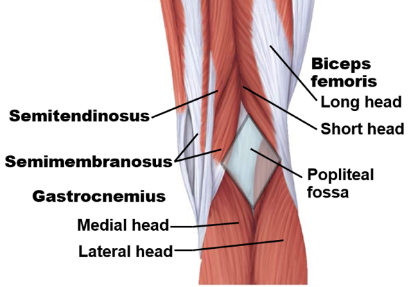 each side and inferiorly by the lateral and medial heads of the gastrocnemius. This region is called the popliteal fossa. When the femoral artery travels through the opening in the adductor magnus and emerges into this space, it is now called the popliteal artery. The popliteal vein also travels with the artery in this space and is continuous with the femoral vein. The popliteal artery ends just inferior to the knee joint as it splits to form the anterior tibial and posterior tibial arteries. The posterior tibial artery may appear as a continuation of the popliteal artery with the anterior tibial artery branching off it.
- Sciatic nerve: The sciatic nerve comprises the tibial and common fibular (peroneal) nerves together within a fibrous sheath. (Note that fibular and peroneal are interchangeable. Peroneal is the older term, and it is slowly being replaced with fibular. Either would be acceptable on an exam.) In the gluteal region, the sciatic nerve runs deep to the piriformis and emerges at the inferior border of this muscle before descending in the posterior thigh. The sciatic does not innervate any muscles as it passes through the thigh, but its branches innervate hamstring muscles. The tibial division innervates the semimembranosus, semitendinosus, and long head of the biceps femoris, while the common fibular division innervates the short head of the biceps femoris. Notice that it splits into the tibial nerve and common fibular (peroneal) nerve in the popliteal fossa. Though this is the most common location of the split, this can occur anywhere along the sciatic nerve path, even as superior as the gluteal region.
- Neurovasculature of the compartments of the leg and foot: The muscles of the leg are divided into four compartments: anterior, lateral, deep posterior, and superficial posterior. Each compartment is typically innervated by one nerve and receives its blood supply from one artery.
- Superficial and deep posterior compartments: The tibial nerve continues into the leg and travels inferiorly between the superficial and deep posterior compartments, innervating the muscles in both compartments. After wrapping around the medial malleolus, it branches into the lateral and medial plantar nerves, which innervate the muscles of the plantar aspect (bottom) of the foot. The posterior tibial artery also travels with the tibial nerve between the superficial and deep posterior compartments, supplying both compartments. It then runs around the medial malleolus and divides into the medial and lateral plantar arteries that supply the medial and lateral aspects of the foot. The lateral plantar artery gives off the plantar arch, which gives off digital arteries to supply the foot and toes.
Anterior, Lateral, and Medial Neurovasculature of the Lower Extremity
- Femoral Triangle: The femoral triangle is a space created by the sartorius laterally, the adductor longus medially, and the inguinal ligament superiorly. The pectineus muscle forms the floor of this space. The femoral nerve, femoral artery, and femoral vein travel through this space wrapped together in a common sheath of tissue.
- Femoral artery: This artery is an extension of the external iliac artery from the pelvis. It is named the femoral artery after it travels deep to the inguinal ligament to enter the leg. Similar to the arteries of the upper extremity, this is a case of the same vessel (“tube”) having a different name depending on its location. This artery supplies the anterior structures of the thigh. Follow this artery as it descends through the thigh, and notice that it travels through an opening in the adductor magnus muscle towards the posterior aspect of the knee.
- The largest branch of the femoral artery is the deep femoral artery. This artery runs posteriorly and supplies the muscles of the posterior and medial regions of the thigh. Also, observe the medial and lateral circumflex femoral arteries branching from the deep femoral artery and traveling towards the femur.
- Femoral nerve: This nerve branches into many small branches shortly after entering the thigh. These branches innervate muscles of the anterior thigh and some fibers of the pectineus, and you can follow the branches to these muscles.
- Femoral vein: This vein travels with the femoral artery through the thigh. It carries blood from the lower extremity back to the pelvis. The artery has a thicker wall and maintains a round shape, while the vein tends to collapse against the artery.
- Femoral artery: This artery is an extension of the external iliac artery from the pelvis. It is named the femoral artery after it travels deep to the inguinal ligament to enter the leg. Similar to the arteries of the upper extremity, this is a case of the same vessel (“tube”) having a different name depending on its location. This artery supplies the anterior structures of the thigh. Follow this artery as it descends through the thigh, and notice that it travels through an opening in the adductor magnus muscle towards the posterior aspect of the knee.
- Anterior compartment of the leg: Observe the common fibular (peroneal) nerve wrapping around the head of the fibula. It then splits into the superficial fibular (peroneal) and deep fibular (peroneal) nerves. The deep fibular nerve travels through the anterior compartment of the leg and innervates the muscles of this compartment. From the popliteal artery and posterior tibial artery, follow the anterior tibial artery as it travels towards the anterior compartment. You can see this artery traveling with the deep fibular nerve deep to the tibialis anterior. As the anterior tibial artery crosses the talocrural (ankle) joint, it becomes the dorsal pedis (dorsalis pedis) artery, which supplies muscles and nerves on the dorsal aspect of the foot. If a clinician suspects blood flow to the foot may be blocked, which could occur with an injury such as a full knee dislocation or occlusion due to atherosclerosis, a clinician will check for a pulse at this artery.
- Lateral compartment: The superficial fibular (peroneal) nerve innervates the muscles of the lateral compartment of the leg. This compartment receives its blood supply from the fibular artery. This artery is a branch of the posterior tibial artery shortly after it branches from the popliteal artery in the popliteal fossa.
- Obturator nerve: Observe the nerve /nerve branches running to adductor longus, adductor brevis, and/or gracilis. These are branches of the obturator nerve, which innervates several muscles in the medial aspect of the thigh.
- Obturator artery: The obturator artery, which branches from the internal iliac artery inferior to the superior gluteal artery, runs anterolaterally along the ilium. It then runs through the obturator foramen of the pelvis along with the obturator nerve.
- The obturator artery gives off the artery to the femoral head. This artery travels through the ligamentum teres and helps to supply the femoral head.
Veins of the Lower Extremity
The veins may not be visible on all of the extremities. Use the Visible Body Atlas or another atlas to view these structures if you cannot identify them on the tissue.
- Observe the two main superficial veins of the leg, the great saphenous vein, and the small saphenous vein. The small saphenous vein drains the lateral foot and leg and drains into the popliteal vein. The great saphenous vein drains the rest of the lower limb and drains into the femoral vein near the hip.
- The deep veins of the lower extremity travel with the artery of the same name. Notice how the anterior tibial vein travels with the anterior tibial artery, and the posterior tibial vein travels with the posterior tibial artery, for example. Furthermore, the fibular vein travels on the posterolateral aspect of the interosseous membrane with the fibular artery. The fibular vein drains the lateral leg and ankle and drains into the posterior tibial vein. The anterior and posterior tibial veins drain the anterior and posterior compartments of the leg, respectively, and unite to form the popliteal vein.
- Moving superiorly, observe the popliteal vein and notice that it ends just superior to the knee. At this point, it becomes the femoral vein. In addition to the great saphenous vein, the deep femoral vein also drains into the proximal portion of the femoral vein. Similar to the femoral artery, the femoral vein becomes the external iliac vein as it passes deep to the inguinal ligament.
Lab Activity 2: Visible Body Digital Atlas-Neurovasculature of the Lower Extremity
We will use the Visible Body Digital Atlas to explore the neurovasculature provided in the Terms to Know. While you are viewing the neurovasculature, use the muscle tables to make connections between the nerves and the muscles that they innervate.
Arteries of the Lower Extremity
Click on the Regions tab, and then choose 10. Hip. In the side Systems toolbar, uncheck the lymphatic, nervous, muscular, and venous systems. You will be left with the bones and arteries of this region. Zoom in on the region of the hip and pelvis. From the anterior view, click on and observe the common iliac artery. This artery originates on each side as the aorta ends and splits in the lower abdominal region. Rotate the image, and observe that the common iliac artery divides into the internal and external iliac arteries.
First, explore the internal iliac artery and its branches. While this artery also supplies the perineum and other pelvic structures, we will focus on the branches that supply muscles of the lower extremity. Click on the superior gluteal artery, a superior branch that runs posteriorly to the gluteal region. With this artery highlighted, rotate the image to a posterior view and observe its path. From the posterior view, click on the internal iliac artery again. Follow this artery inferiorly, and, as it divides, click on the more lateral artery, the inferior gluteal artery. While you still have the inferior gluteal artery highlighted, click on the radius blast (bullseye) three times. Observe how the ligaments of the pelvis form the greater sciatic foramen in the region of the greater sciatic notch of the pelvis, and notice how these arteries both travel through the greater sciatic foramen. The piriformis muscle has also been added to this image. You can now see that the superior gluteal artery leaves the greater sciatic foramen superior to the piriformis muscle, while the inferior gluteal artery leaves the greater sciatic foramen inferior to the piriformis muscle. The superior gluteal artery supplies the gluteus maximus, gluteus medius, gluteus minimus, and tensor fasciae latae muscles, while the inferior gluteal artery supplies the gluteus maximus, two lateral rotators, and the superior parts of the hamstring muscles.
Return to the anterior view, where you started this section, and remove the lymphatic, nervous, muscular, and venous systems. Now click on the obturator artery, which branches from the internal iliac artery just inferior to the superior gluteal artery and runs anterolaterally along the ilium. Observe how this artery runs through the obturator foramen of the pelvis. With this artery highlighted, rotate the image to view it from different angles. It supplies the thigh adductors, the proximal part of the hamstrings, and one lateral rotator muscle. The obturator artery gives off an anterior and a posterior branch, though you are not responsible for identifying these branches. However, you should know that the posterior branch of the obturator artery gives off the acetabular branch or the artery to the femoral head. Though it is not visible on the iPads, you should know that this artery travels through the ligamentum teres and helps to supply the femoral head.
Zoom out to view the full hip and thigh. Just distal to the common iliac artery, now click on the external iliac artery. Branches of this artery supply the rest of the lower extremity. Notice that the external iliac artery ends as it passes behind the inguinal ligament. Distal to the inguinal ligament, this vessel becomes the femoral artery. Zoom in to take a closer look at the branches of the femoral artery. Observe the deep femoral artery running posteriorly. The deep femoral artery gives off two branches that supply the hip joint: The medial and lateral circumflex femoral arteries. They form an anastomosis to supply the hip region and the head and neck of the femur. Rotate the image to observe how this artery wraps around the proximal femur.
Observe the femoral artery from a medial view and notice that it moves posteriorly as it descends in the medial thigh. Also, notice that the femoral artery ends just superior to the knee joint. At this joint, the artery  becomes known as the popliteal artery. This artery will be best viewed posteriorly.
The popliteal artery ends just inferior to the knee joint as it splits to form the anterior tibial and posterior tibial arteries.
Rotate the image to an anterior view, click on the anterior tibial artery, and observe that it passes through the interosseous membrane. It descends on its anterior aspect, supplying the anterior compartment of the leg. As it crosses the talocrural joint, it becomes the dorsal pedis artery.
The posterior tibial artery supplies both the deep and superficial posterior compartments. Soon after branching off the popliteal artery, it gives off the fibular artery, which travels just posterior to the interosseus membrane. The fibular artery supplies the lateral compartment of the leg.
Look at the plantar aspect of the foot. Observe the posterior tibial artery running around the medial malleolus and then dividing into the medial and lateral plantar arteries (you will need to hide the plantar fascia to see this). Also, observe the plantar arch and digital arteries.
Veins of the Lower Extremity
Click on the Regions tab, and then choose 10. Hip. In the side Systems toolbar, uncheck the lymphatic system, nervous system, and muscular system. You will be left with the bones, arteries, and veins of this region. Zoom in on the region of the hip and pelvis. First, observe the two main superficial veins of the leg, the great saphenous vein, and the small saphenous vein. The small saphenous vein drains the lateral foot and leg and drains into the popliteal vein. The great saphenous vein drains the rest of the lower limb and drains into the femoral vein near the hip.
The deep veins of the lower extremity travel with the artery of the same name. Notice how the anterior tibial vein travels with the anterior tibial artery, and the posterior tibial vein travels with the posterior tibial artery. Furthermore, the fibular vein travels on the posterolateral aspect of the interosseous membrane with the fibular artery. The fibular vein drains the lateral leg and ankle and drains into the posterior tibial vein. The anterior and posterior tibial veins drain the anterior and posterior compartments of the leg, respectively, and unite to form the popliteal vein.
Moving superiorly, observe the popliteal vein and notice that it ends just superior to the knee. At this point, it becomes the femoral vein. In addition to the great saphenous vein, the deep femoral vein also drains into the proximal portion of the femoral vein. Similar to the femoral artery, the femoral vein becomes the external iliac vein as it passes deep to the inguinal ligament.
Nerves of the Lower Extremity
Click on the Regions tab, then 10. Hip. In the side Systems toolbar, uncheck the lymphatic system, muscular system, and the vascular systems. You will be left with the bones and nerves of this region. Zoom in on the region of the hip and pelvis from a posterior view. Now use the Anatomy Search icon and type in piriformis. Add the piriformis as well as the gluteus minimus. Similar to the arteries of the same name, observe that the superior gluteal nerve leaves the greater sciatic foramen superior to the piriformis, while the inferior gluteal nerve leaves the greater sciatic foramen inferior to the piriformis. The superior gluteal nerve innervates the gluteus medius, gluteus minimus, and tensor fasciae latae muscles, while the inferior gluteal nerve innervates the gluteus maximus only.
Move to an anterior view. Click on the femoral nerve, and observe its branches traveling to the muscles of the anterior thigh. Now rotate to a posterior view. Observe a nerve running through the obturator foramen. This is the obturator nerve, and it innervates the muscles of the medial thigh (except the hamstring portion of the adductor magnus).
In this view, you can also observe the sciatic nerve exiting through the greater sciatic foramen inferior to the piriformis. As you move inferiorly, you will observe that the tibial and common fibular nerves comprising the sciatic nerve separate from each other in the superior popliteal fossa.
Observe the tibial nerve traveling through the posterior leg and branching into the lateral and medial plantar nerves (you will need to hide the plantar fascia to see this). Also, observe the common fibular (peroneal) nerve running rather superficially, wrapping around the head of the fibula. Then it splits into the superficial and deep fibular (peroneal) nerves. Notice that the deep fibular (peroneal) nerve travels just anterior to the interosseous membrane. This nerve innervates the muscles of the anterior compartment of the leg and the dorsal aspect of the foot. The superficial fibular (peroneal) nerve innervates the muscles of the lateral compartment of the leg.
