
Lab 6: The Cerebrum
Learning Objectives:
- Explain the directional terms associated with the orientation of the brain in the cranial cavity.
- Describe the layers of tissue that cover the brain and explain their function.
- Describe the structures of the cerebral hemispheres, including the lobs of the brain and their components, using the whole-brain and midsagittally sectioned tissue.
- Explain the various functions associated with identified regions of the brain.
- Describe the Circle of Willis.
Terms to Know
|
Directional Terms
Cerebrum
|
Meninges
Ventricles
Other Structures
Blood Supply to the Brain
|
Introduction
In today’s lab, you will learn about the cerebrum of the brain and introduce the brainstem and cerebellum. This mass of neurons and glial cells controls everything from our ability to breathe, speak, and move our limbs, to our personalities, emotions, and higher-level thinking.
You will use various tools in this lab, including the Navigator, ventricle models, and, most importantly, human brain tissue. The brain tissue must be handled with care and respect. Take extra care to ensure that these brains are covered with towels when they are not in use. Also, handle the tissue carefully to ensure you do not gouge or nick the brain surfaces or pull excessively at parts of the tissue.
Before we begin with the lab activities, it is essential to address additional directional terms used with respect to the brain. A change in the long axis of the nervous system occurs between the cerebrum and the brainstem. The long axis of the cerebrum is relatively horizontal, while the long axis of the brainstem and spinal cord is relatively vertical.
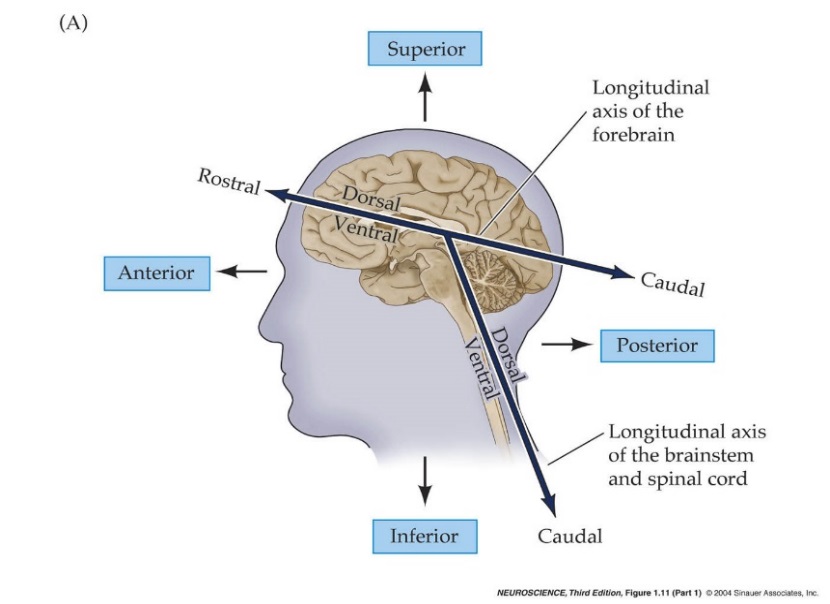
As a result, some of the directional terms may have slightly different meanings depending on the structure to which they refer. Rostral refers to the most frontal portion of the brain (rostrum = nose), while caudal describes structures closer to the tip of the spinal cord (caudal = tail). When used in the context of the cerebrum, rostral refers to structures closer to the nose (as before), but caudal now refers to structures toward the back of the head (posterior). The terms ventral and dorsal can be used interchangeably with anterior and posterior with respect to the plan of the brainstem and spinal cord. However, in the plane of the cerebrum, these terms are interchangeable with inferior and superior.
The University of British Columbia has excellent resources that you may find helpful in studying the brain. These resources can be found at neuroanatomy.ca. The site contains videos, interactive modules, 3D models, cross-sectional images, radiology, and other tools.
Lab Activity 1: Human Brain Tissue
We have several brains available for you to learn from in the lab. Some are whole brains, some are cut midsagittally and divided into hemispheres, and others have been sectioned coronally. As you will observe, they all have the same structures, but there is some variation between each anatomically.
The Meninges
First, explore the meninges. Three connective tissue membranes cover the brain: dura mater, arachnoid mater, and pia mater. The brain has a consistency like somewhat firm jelly during life. The meninges protect this soft structure by anchoring it to the skull and preventing excessive movement within the skull.
The dura mater has been removed from the brain tissue, but it is present in one of the brain cases. Observe how thick this meningeal layer is. The dura mater follows the contours of the skull’s inner surface and does not dive into the sulci of the brain. In a few places, the dura mater folds and dives into spaces between parts of the brain. These are called the dural reflections. You can explore the dural reflections in the images below.
The arachnoid mater can be seen on the surface of some of the brain tissue. It is the middle layer of the meninges, and it is a delicate, transparent membrane. The arachnoid mater does not follow the contours of the sulci and gyri; instead, it follows the form of the overlying dura. On several of these brains, you can notice aggregations of tiny white granules near the superior midline of the brain. These are arachnoid granulations. They function to return cerebrospinal fluid from the subarachnoid space (between this arachnoid mater layer and the pia mater layer of meninges) to the blood.
The pia mater is the innermost dural membrane. It cannot be seen with gross examination of the brain, but it covers the surface of the brain tissue, including within the sulci and gyri, of these brains.
Cerebral Hemispheres: Gross Examination
The cerebral hemispheres contain the brain regions involved in our higher cognitive functions, including language, learning, memory, and personality. The surface of the cerebral hemispheres is made up of the cerebral cortex, which is a layer of gray matter. This surface is thrown into many folds forming sulci and gyri. The sulci are the folds diving inward, while the gyri are ridges of the cortex that are visible on the surface of the brain. Sulci and gyri are essential because they increase the surface area of the cortex, giving us more room for neurons involved in higher cognitive functions.
Use the whole-brain and midsagittally sectioned brain tissue to identify the following structures of the cerebral hemispheres.
- The median longitudinal fissure separates the left and right hemispheres.
- The corpus callosum is the large white matter pathway connecting the right and left hemispheres of the brain. Observe this structure in cross-section on a midsagittally sectioned tissue.
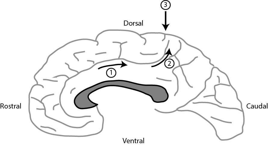 The central sulcus marks the boundary between the frontal and parietal lobes. It can be tricky to identify, so to reliably identify it, start from the midsagittal section. Above the corpus callosum (gray in the image) is a curved gyrus called the cingulate gyrus. The sulcus dorsal to the gyrus is the cingulate sulcus. If you follow the cingulate sulcus (1) in the direction of the arrows, it bends dorsally (2) and comes to a stop very close to the dorsal aspect of the brain surface. Move rostrally one sulcus, and you’ve located the central sulcus (3). Now you can trace it over the lateral aspect of the brain.
- The precentral gyrus is in the frontal lobe, just anterior to the central sulcus. This is where the primary motor cortex is located, where all voluntary motor signals begin.
- The postcentral gyrus is in the parietal lobe just posterior to the central sulcus. This is the primary somatosensory cortex, which processes general sensory information.
- The parieto-occipital sulcus separates the parietal lobe from the occipital lobe.
- The calcarine sulcus (fissure) is in the occipital lobe. It runs roughly perpendicular to the parieto-occipital sulcus and contains the primary visual cortex. These are visible on the midsagittal section.
- The lateral (Sylvian) fissure is the boundary between the frontal & temporal lobes.
- The temporal lobe contains three gyri parallel to the lateral (Sylvian) fissure: the superior temporal gyrus, middle temporal gyrus, and inferior temporal gyrus. The primary auditory cortex is located in the superior temporal gyrus.
- The insula is located deep within the Sylvian fissure. Its functions are not well understood, but taste is one function that it is thought to play a role in. It may be difficult to see on the whole brain, so be sure to look at it in cross-section as well.
- The thalamus is located deep in the brain and cannot be seen in the whole-brain tissue. This is the relay center of the brain. Many pathways between the cerebrum, brainstem, cerebellum, and spinal cord have a synapse in the thalamus.
- The hypothalamus is located just anterior and inferior to the thalamus. This structure is responsible for many functions, including endocrine control, species-preserving behaviors (hunger, thirst), and circadian rhythm.
- The anterior commissure is located just anterior to the thalamus. It connects parts of the frontal and temporal lobes of the two hemispheres.
Though not part of the cerebrum, observe the cerebellum and the three parts of the brainstem: the midbrain, pons, and medulla oblongata. You can only see the midbrain in the midsagittal section, as it is mostly hidden by other structures in the whole brain tissue. For now, be familiar with the general location of these structures. We will discuss them more in-depth in the next lab.
The Circle of Willis: Blood Supply to the Brain
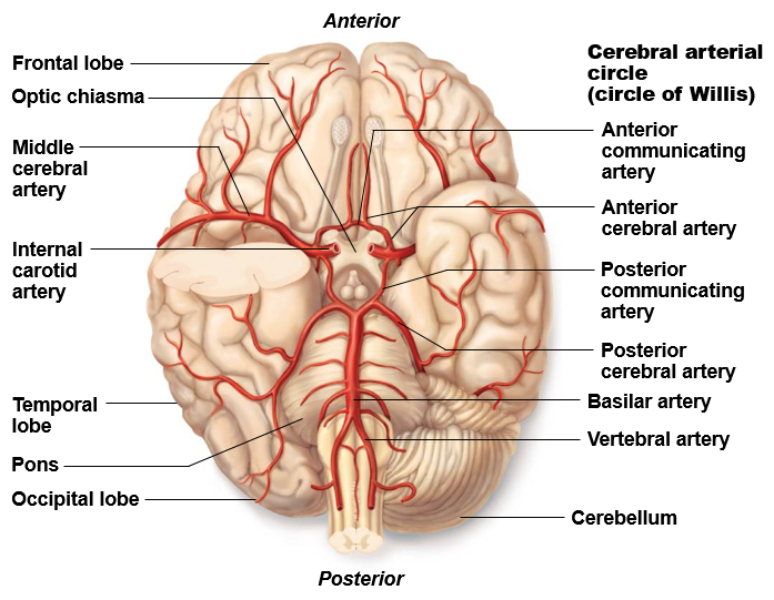
The brain is supplied by two pairs of arteries: the vertebral arteries and the internal carotid arteries. They branch into arteries that eventually supply the whole brain. First, look at these arteries and their branches on the inferior aspect of the whole-brain tissue. Then observe the anterior cerebral artery on the midsagittal section.
- The vertebral arteries merge along the brainstem to form the single basilar artery.
- The basilar artery travels along the ventral surface of the pons (part of the brainstem). It gives off several branches to the cerebellum along its path.
- The basilar artery splits on the ventral surface of the pons to give off right and left posterior cerebral arteries. The posterior cerebral arteries supply the posterior aspect of the brain, including the occipital lobes, as well as the inferior portion of the temporal lobes.
- Locate the internal carotid arteries. They travel through the skull and have been cut here, so it looks like their lumen is facing you.
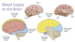
- Each internal carotid artery divides into two branches: the anterior cerebral artery and the middle cerebral artery. The middle cerebral artery dives into the lateral fissure to travel to the lateral aspect of the brain, where it supplies the majority of the temporal lobe and a large portion of the frontal and parietal lobes (lateral aspects). The anterior cerebral artery travels anteromedially to the median longitudinal fissure where it supplies the superior and medial aspects of the frontal and parietal lobes. Examine a midsagittal section to follow the anterior cerebral artery as it runs along the anterior and superior border of the corpus callosum.
- The two anterior cerebral arteries are connected just before they enter the median longitudinal fissure by the anterior communicating artery. This is usually a very short artery connecting them, or the two anterior cerebral arteries can appear to “touch” each other and then split again.
- The posterior communicating arteries are small arteries that connect the posterior cerebral and internal carotid arteries.
The three pairs of cerebral arteries and the three communicating arteries form the Circle of Willis, an arterial circle that provides collateral circulation. There is a great deal of variation in the sizes of the component arteries of the circle, including instances of left-right asymmetry. It is not unusual to have an incomplete circle of Willis, and you may notice some variations in the brain tissue in our lab.
Activity 2: Ventricle Models and the Flow of Cerebrospinal Fluid
The brain contains several open spaces, called ventricles, filled with cerebrospinal fluid. The ventricle models represent the shape of the open, fluid-filled spaces in the brain. Utilize both the ventricle models and obtain a midsagittally sectioned brain tissue to see the ventricles. The models have laminated images and a key.
Observe the open spaces in the middle of the hemisphere. These are the lateral ventricles. The septum pellucidum is a thin membrane that separates the anterior part of the lateral ventricles. The larger anterior portion is called the anterior horn of the lateral ventricle. The body of the lateral ventricle is the thinner portion, just posterior to the anterior horn. The portion in the temporal lobe is called the inferior horn of the lateral ventricle. The posterior horn of the lateral ventricle extends posteriorly.
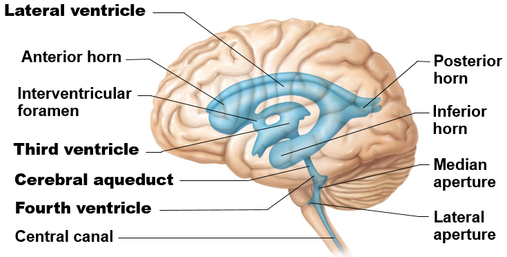
The third ventricle is a thin midline space that separates the left and right thalami. Each lateral ventricle connects to the third ventricle by way of the interventricular foramen (of Monro). Notice that the third ventricle in the image to the right appears to have a hole in the middle of it. This is created by a midline thalamic structure called the massa intermedia, or interthalamic adhesion, which connects the two thalami and passes through the third ventricle.
The third ventricle connects to the fourth ventricle via the cerebral aqueduct. The fourth ventricle lies between the pons and the cerebellum.
All of the ventricles contain choroid plexus, which produces cerebrospinal fluid (CSF) within the ventricles. This can be observed within some of the ventricles in the midsagittally sectioned tissue & the coronal sections. The choroid plexus can produce CSF in all cerebral ventricles, but the longest possible pathway of CSF flow begins in the lateral ventricle. Locate the midsagittally sectioned brain and the ventricle model to follow the flow of CSF from the production in the choroid plexus in the lateral ventricle through the CNS.
- Lateral ventricle–>Interventricular foramen (of Monro)–>Third ventricle–>Cerebral aqueduct–>Fourth ventricle–>From the fourth ventricle, CSF can escape via the lateral apertures, median aperture, or central canal. These apertures can only be observed on the model.
Lab Activity 3: Visible Body App
Observe the following structures in the Visible Body app:
- Meninges: To see the meninges go to the search button and type in meninges. Here you can click on the outer layer of the meninges, the dura mater. If you hide the dura mater, you will be able to see the falx cerebri and the tentorium cerbelli. **You cannot see the arachnoid or pia mater in the app.
- Cerebrum: To see the cerebrum and associated structures, under the Nervous System Views, click on 2. Brain. You will have to hide some of the skull bones. Still, you will be able to see each left cerebral hemisphere, sulci and gyri, corpus callosum, each lobe, precentral gyrus, postcentral gyrus, central sulcus, lateral sulcus, parieto-occipital sulcus, basal ganglia, caudate nucleus, and putamen if you go back and click on 5. Thalamus, you will be able to see the thalamus and hypothalamus.
- Ventricles: To see the ventricles, under the Nervous System Views click 4. Limbic System. You will see the lateral ventricle, third ventricle, cerebral aqueduct, and fourth ventricle from this view.
- Blood Supply to the Brain: To see the arteries of the brain, under the Circulatory System Views, click on 4. Circle of Willis. You will see vertebral arteries, internal carotid arteries, anterior cerebral arteries, middle cerebral arteries, posterior cerebral arteries, anterior communicating artery, and posterior communicating artery.
Lab Activity 4: Cross-sectional Anatomy of the Brain
The Navigator
Observe cross-sectional images of the brain using the Navigator. Be sure to observe slices throughout the brain. Many structures span a large portion of the brain, while others are only visible in a few slices.
Observe the following structures in cross-section:
- Median longitudinal fissure: Space between the two hemispheres superiorly.
- Corpus callosum: This can be observed as the thick structure connecting the two hemispheres just inferior to the median longitudinal fissure.
- Sylvian fissure: Lateral space between the temporal lobe and the frontal or parietal lobes.
- Insula: Observed laterally but deep, buried within the depths of the Sylvian fissure.
- Anterior commissure: This commissural pathway (connecting the two hemispheres) is only visible for a few slices. It is observed connecting the hemispheres inferiorly.
- Lateral ventricles: The anterior horn, body, and posterior horn appear in the center of each hemisphere anteriorly, in slices towards the middle, and posteriorly in the brain. The inferior horn can be seen next to the hippocampus in the temporal lobe, and it is typically relatively thin and flat in shape.
- Third ventricle: Located between the right and left thalami (thalami = plural of thalamus).
- Amygdala: Located in the anterior portion of the medial temporal lobe. This appears as a center of gray matter in this region. It is involved in behavior and giving emotional meaning to sensory input and memory.
- Hippocampus: Observed just posterior to the amygdala in the medial temporal lobe. While the amygdala is a region of gray matter, the hippocampus appears curled next to the inferior horn of the lateral ventricle. The hippocampus is essential for storing new memories and is often atrophied in Alzheimer’s disease.
- Caudate nucleus: Located deep in the brain next to the lateral ventricle. It is larger anteriorly and smaller posteriorly and somewhat C-shaped in the sagittal plane. The caudate is part of the basal ganglia, a collection of nuclei involved in the control of voluntary motor activity.
- Internal capsule: This white matter pathway is lateral to the caudate nucleus, between the caudate and the putamen (and globus pallidus). It carries primarily motor fibers, including corticospinal tract fibers.
- Globus pallidus and Putamen: These are located just lateral to the internal capsule. The putamen is lateral and slightly superior to the smaller globus pallidus. The putamen is visible alone more anteriorly, and both structures are visible near the middle of the brain.
- Thalamus: Consists of a collection of nuclei that sit posterior to the basal ganglia structures. You do not need to know the different nuclei of the thalamus, but you will be able to observe some of the different nuclei in cross-section.
- Hypothalamus: Located just inferior to the thalamus.
Coronal Brain Cross Sections
Observe the above structures in the coronally-sectioned brain tissue. When observing the medial temporal lobe, notice the significant atrophy of both the amygdala and hippocampus. This may be due to neurodegenerative disease, but we do not know the cause of death or medical history for this individual. If you observe the corresponding midsagittally sectioned hemisphere, you can see the atrophy of the intact medial temporal lobe.
Be sure to observe all coronal sections throughout the brain. Some structures are only visible in the anterior sections, while others are only visible posteriorly.
When observing the color-coded images included at this station, note that the brain sections are stained so that the white matter appears very dark compared to the gray matter.
Lab Activity 5: Radiology of the Brain
Use the radiology paired with the University of British Columbia cross-sections page by clicking on “View MRI” or “View Labelled MRI” in the column at the right. Not all slices have associated radiology images, but this tool will allow you to view radiology images of the brain alongside cross-sectional images of the same slice.
