
Lab 7: Brainstem, Cranial Nerves, and Skull
Learning Objectives:
- Describe the anatomical features of the cerebellum and brainstem.
- Identify cranial nerves I-XII.
- Describe the anatomy of the skull and identify the bones of the skull.
- Identify the selected foramen of the skull & explain the structures that run through these foramen.
Terms to Know
|
Cerebellum
Brainstem
Cranial Nerves
|
Skull
|
Introduction
We continue discussing the brain by examining the brainstem, cerebellum, and cranial nerves in this lab. We will also explore the bones, landmarks, fossa, and sutures of the skull. We combine the cranial nerves and skull into one lab because the cranial nerves will travel through the foramina of the skull that you will learn about. This material lends itself well to methodical and regular studying, and be sure to study the foramina and cranial nerves that travel through them together.
Lab Activity 1: Brain Tissue – Brainstem, Cerebellum, Cranial Nerves
Observe the brainstem, cerebellum, and cranial nerves on the brain tissue. Also, use the atlases and iPad app to help you find each structure on the tissue. If you are having trouble identifying something, please ask a TA for help.
**Please remember to be GENTLE with the brain tissue. Do not dig around, spread apart lobes, or press down with more than a light touch on the brains. Treat them with care, as we will be using these same brains for several years.
Cerebellum
The cerebellum coordinates voluntary movements and helps to maintain posture and equilibrium. It receives information from the primary motor cortex of the frontal lobe about the intended movement, compares this intended movement with the actual positioning and movement of the body, and sends signals back to the frontal lobe to resolve any differences between the intended movement and current position. The cerebellum is also involved in coordinating higher cognitive functions and emotions.
On the whole brain tissue, locate the cerebellum and observe the two hemispheres. Also, observe the numerous fine grooves, or sulci, on the surface of the cerebellum running transversely. These are called the folia, and they are similar to the gyri of the cerebrum.
Observe the folia on the midsagittally sectioned brain. Also, observe how the fourth ventricle separates the cerebellum from the pons of the brainstem.
Brainstem
The three major regions of the brainstem, from inferior to superior, are the medulla oblongata, the pons, and the midbrain. The brainstem contains cranial nerve nuclei (collections of neurons) that communicate with the cerebrum, cerebellum, and spinal cord, as well as nuclei that form the cranial nerves. It also contains sensory and motor pathways connecting the cerebrum and the spinal cord.
Locate the whole brain and midsagittally section brain tissue. Use these to identify the following major brainstem structures.
- Locate the pons (bridge) on the ventral surface of the brainstem. The pons can be easily identified by its anterior bulge.
- Rostral to the pons, observe the midbrain. It is located just inferior to the thalamus, which is best viewed in the midsagittal section. The temporal lobes somewhat hide this structure. Observe the following structures of the midbrain:
- The midbrain surrounds the cerebral aqueduct, which is the pathway that CSF travels between the third and fourth ventricles. This is best seen in a midsagittal view.
- The cerebral peduncles are located on the ventral side of the midbrain. These white matter pathways carry fibers of the corticospinal tract, originating in the primary motor cortex and descending fibers from other cortical regions. The space between the cerebral peduncles is called the interpeduncular fossa.
- On the midsagittal tissue without the brainstem, look at the cut brainstem and observe a dark pigmented line in the midbrain. This is the substantia nigra. In Parkinson’s disease, the neurons in this structure are destroyed, and the structure is light or no longer visible.
- Observe the corpora quadrigemina, also known as the tectum. This is best seen on one of the cross section cut brain specimens. It appears as four bumps on the posterior (dorsal) surface of the midbrain.
- The two superior bumps are called the superior colliculi. They are involved in visual reflexes, causing you to orient your head to focus your eyes on something seen in your periphery.
- The two inferior bumps are called the inferior colliculi. They are involved in auditory reflexes, causing you to orient your head to focus your eyes on something you hear.
- Caudal to the pons, examine the medulla oblongata. The medulla oblongata is continuous with the spinal cord. Identify the following on the medulla oblongata:
- Locate the pyramids, the eminences of the corticospinal (pyramidal) tract that appear as tube-like bulges on the ventral aspect of the medulla oblongata. The pyramids are visible from their emergence at the pons to the pyramidal decussation at the junction between the medulla oblongata and spinal cord. Identify the pyramidal decussation where the sulcus between them disappears briefly. This is the decussation of the fibers of the corticospinal tract.
Cranial Nerves
The cranial nerves are peripheral nerves that carry various sensory and/or motor information from/to the head and neck. Ten of the twelve cranial nerves are associated with the brainstem, while cranial nerves I (olfactory) and II (optic) are associated with the cerebrum and thalamus, respectively. The functions of each of these nerves are described here. As part of these descriptions, we will mention structures that you haven’t learned yet. However, we will discuss all of these structures by the end of this unit.
You can observe the cranial nerves on the whole-brain tissue. Some of the cranial nerves are not present on some of the brains or some sides (right or left) of the brains. However, between all whole brains, atlas images, and the Visible Body app, you should be able to observe all cranial nerves.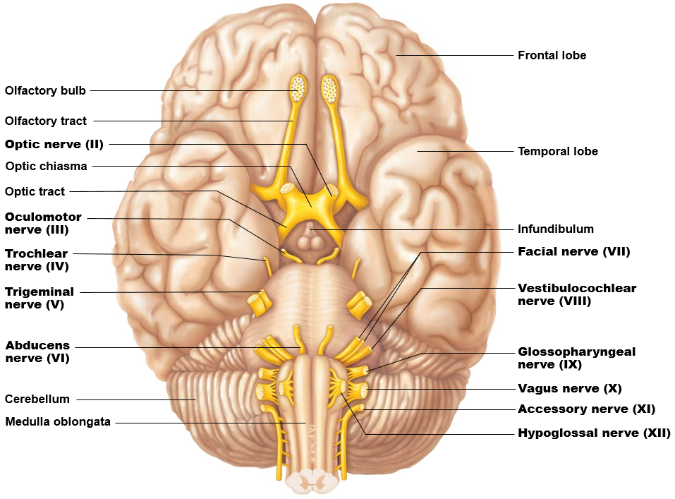
- Olfactory Bulbs and Tracts – CN I: The olfactory nerves arise in the olfactory epithelium of the nasal cavity and course dorsally to the olfactory bulb inferior to the frontal lobe of the cerebral hemisphere. The actual nerves were lost during the removal of the brain. Observe the olfactory bulb, where these nerves synapse, on the inferior aspect of the frontal lobe. The olfactory tract originates in the olfactory bulb and runs caudally on the ventral aspect of the frontal lobe.
- Optic Nerves – CN II: The optic nerves begin in the retina of each eye. These nerves course posteriorly and are united in the optic chiasm. The fibers split again immediately posterior to the optic chiasm and extend posteriorly as the optic tracts.
- Oculomotor Nerves – CN III: These motor nerves emerge in the interpeduncular fossa and innervate 4 of the 6 extraocular muscles: the medial rectus, superior rectus, inferior rectus, and inferior oblique.
- Trochlear Nerves – CN IV: These are the only nerves to emerge on the brainstem’s dorsal aspect. They travel around the sides of the midbrain and pons to innervate the superior oblique. You can find these tiny nerves between the lateral rostral pons and the cerebrum.
- Trigeminal Nerves – CN V: These large nerves are the only cranial nerves to emerge from the lateral aspect of the pons. This nerve innervates the muscles of mastication (chewing) and conveys somatic sensation from the entire face.
- Abducens Nerves – CN VI: These nerves emerge near the midline at the border of the pons and the medulla oblongata. They innervate the lateral rectus muscle of the eye.
- Facial Nerves – CN VII: These mixed motor and sensory nerves emerge from the junction of the pons and medulla oblongata lateral to the emergence of the abducens nerves. They carry sensory information from the taste buds of the anterior two-thirds of the tongue and innervate the muscles of facial expression, the lacrimal glands of the eye, and most salivary glands.
- Vestibulocochlear Nerves – CN VIII: These nerves, which carry both auditory and vestibular sensation, enter the brainstem lateral to (right next to) the facial nerves.
- Glossopharyngeal Nerves – CN IX: These nerves are composed of the most rostral of a series of rootlets that emerge posterior to the olive on the lateral aspect of the medulla oblongata. They receive sensory information from the tonsils, pharynx, middle ear, and posterior tongue and innervates the stylopharyngeus muscle and parotid gland.
- Vagus Nerves – CN X: These nerves emerge caudal to the glossopharyngeal nerves in the same series of rootlets. The vagus nerve has several functions, including providing parasympathetic innervation to organs of the thorax and part of the abdomen and the innervation of muscles of the larynx and pharynx.
- Spinal Accessory Nerves – CN XI: These nerves, which innervate the trapezius and sternocleidomastoid muscles, emerge caudal to the vagus nerve.
- Hypoglossal Nerves – CN XII: These motor nerves innervate the musculature of the tongue. They emerge from the ventral aspect of the caudal medulla as a series of rootlets in the groove just lateral to the pyramids.
Tip: The cranial nerves with only motor functions (oculomotor, trochlear, abducens, and hypoglossal) emerge medially compared to the other cranial nerves. You cannot see this with the trochlear nerve because it emerges on the brainstem’s dorsal aspect, but you can notice this with CN III, CN IV, CN VI, and XII.
Review the summary of cranial nerves table. The chart describes the cranial nerves, their passageways through the skull, and their functions.
Lab Activity 2: Visible Body Atlas- Cerebellum, Brainstem, Cranial nerves
Use the iPads to identify the structures of the cerebellum, brainstem, and cranial nerves. Under the Nervous System Views, click on 2. Brain, 5. Thalamus, and 6. Cranial Nerves. Within these views, you will be able to see the cerebellum, medulla oblongata, pons, midbrain, and all of the cranial nerves. **Note** you will not be able to see the associated structures for the cerebellum, medulla oblongata, or midbrain in the app. You will need to hide layers of the skull, zoom in/out and rotate the image to see all structures.
Use the iPads to identify the structures of the skull. Open the atlas app on the iPad, and under Skeletal System Views, click 2. Skull. Here you will be able to see the sutures from the list above. However, they are not identified in the app. Return to the menu and click 3. Cranial Fossae. Here you will be able to see the fossa listed above. However, they are not identified in the app. While in this view, you will be able to identify and see CN I-XII. You will have to zoom in/out and rotate the image to see all the cranial nerves. While manipulating the image, you should also identify and see the passageways for each cranial nerve (**see table at the end of the lab guide to help determine which passageways to look for). To click on the passageways, you will need to zoom in and specifically touch the passageway. You will identify every bone and listed anatomical feature from the terms above while in views 2. Skull, 3. Cranial Fossae, 4. Skull, Sagittal Section, 5. Skull Coronal Section and 6. Disarticulated Skull (this view particularly will allow you to see every bone of the skull).
Lab Activity 3: Navigator – Brainstem, Cerebellum, Cranial Nerves
Use the Navigator to observe the cerebellum, brainstem, and skull. You can click on Advanced to select/unselect structures and isolate the brainstem and spinal cord or isolate specific cranial nerves to observe where they emerge from the brainstem or travel through the skull.
You can also review the cross-sectional anatomy of the brain covered in the previous lab while you are at the Navigator in this lab.
Lab Activity 4: Skull – Bones and Foramen
Use the human skulls to identify the bones, major features, and passageways of the skull. Use a paper or digital atlas to help you identify the bones and passageways listed below. Also, be sure you have pipe cleaners, as you will use these to traverse several of the passageways of the skull.
- First, observe the bones that make up the majority of the external aspect of the cranium: the parietal bones, occipital bone, temporal bones, and frontal bone.
- On the temporal bone, identify the mastoid process and styloid process. These are important sites of muscle attachment.
- Look at the inferior aspect of the skull. Observe the occipital condyles at the inferior portion of the occipital bone, which makes up most of the inferior portion of the cranium. These condyles articulate with the atlas of the vertebral column.
- Observe the places where these bones meet. These are called sutures.
- Coronal suture: where the frontal bone meets the two parietal bones.
- Sagittal suture: where the right and left parietal bones meet.
- Lambdoid suture: between the parietal bones and occipital bone.
- Squamous suture: between the parietal bone and temporal bone on each side.
- Then observe the bones that make up the face.
- The frontal bone is the bone of the forehead.
- The zygomatic bone makes up the anterior cheek. The prominent part of the zygomatic bone meets part of the temporal bone laterally to form the zygomatic arch, which makes up our cheekbone.
- The nasal bone forms the superior portion of the nose.
- The maxilla forms the upper jaw and medial portion of the cheek.
- The mandible forms the lower jaw. This is the part of the jaw that moves when we chew or speak. The anterior tip of the mandible that forms the chin is called the mental protuberance. Also, observe the articulation between the mandible and the temporal bone. This is the temporomandibular joint. The posterior structure that articulates with the temporal bone is the condylar process. The flat portion protruding up just anterior to this is the coronoid process of the mandible.
- Remove the top of the cranium from the skull. There are a few bones that are best viewed from the internal aspect of the cranium.
- First, observe that bony landmarks within the skull form three bowl-like spaces. These spaces are called the anterior cranial fossa, middle cranial fossa, and posterior cranial fossa.
- The ethmoid bone is a small anterior, medial bone located within the anterior cranial fossa. You can identify it by the several small holes in it.
- Just posterior to the frontal and ethmoid bones, observe the sphenoid bone. This bone has a ridge that separates the anterior cranial fossa from the middle cranial fossa. The temporal lobes of the brain sit in the middle cranial fossa.
- The rest of the middle cranial fossa is made up of the temporal bone. The posterior aspect of the temporal bone has a ridge that separates the middle cranial fossa from the posterior cranial fossa. The posterior cranial fossa is made up of the occipital bone, and this space holds the cerebellum.
There are additional bones of the skull and additional features of these bones that we will discuss in this unit, such as the orbit, nasal cavity, and oral cavity. However, we will discuss these bones and features with other labs.
Many nerves and vessels enter and leave the skull. Therefore, there are many passageways within the skull. You will be asked to identify several of these passageways. Additionally, you will be asked to know which nerves or vessels travel through these passageways.
Using the atlas and a skull, identify the passageways listed in the chart below. Pass a pipe cleaner through the larger passageways to see where they exit the skull. Note that the stylomastoid foramen cannot be observed on the internal aspect of the skull. This foramen is visible between the mastoid process and styloid process of the temporal bone.
| Foramina/Passageways | Contents (items that pass through) |
| Anterior Cranial Fossa | |
| Cribriform foramina in cribriform plate | Olfactory nerves (CN I) |
| Middle Cranial Fossa | |
| Optic canals | Optic nerves (CN II) and ophthalmic arteries |
| Superior orbital fissure | CN III, CNIV, & CN VI; (Ophthalmic) branch of CN V |
| Foramen ovale | (Mandibular) branch of CN V nerve |
| Carotid canal | Internal carotid artery |
| Posterior Cranial Fossa | |
| Foramen magnum | Medulla oblongata and meninges, vertebral arteries** |
| Jugular foramen | CN IX, X, and XI; internal jugular vein |
| Hypoglossal canal | Hypoglossal nerve (CN XII) |
| Internal acoustic meatus | CN VII and CN VIII |
*Note: The trigeminal nerve gets its name because it divides into three branches (the name means “three twins”). The different branches pass through different foramina of the skull. You do not need to know or identify the branches of the nerve (in parentheses in the chart) in this lab. You will just be asked to know that a branch of the trigeminal travels through the specific foramina above.
**A portion of the accessory nerve does travel through foramen magnum, but we will not test you on that in this course.
