
M4 | Descending control – Lateral system
Primary motor cortex
Organizational features
Somatotopy
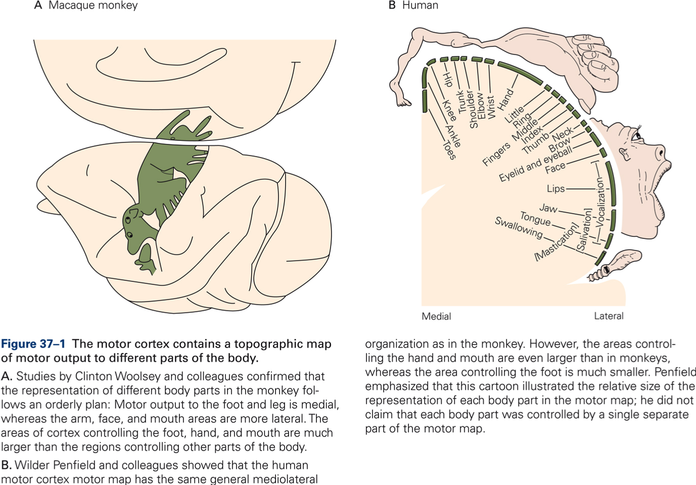
- Electrical stimulation evokes movements of specific parts of the body.
- Thresholds for eliciting movements are lowest in MI, they are higher in SMA and PM.
- Movements more complex in SMA and PM than in MI (e.g. orienting body, opening/closing hand).
- Movements of adjacent parts of the body controlled by neurons in adjacent parts of a structure. For example, within MI the leg is represented medial, the arm more lateral, and the face most lateral.
- More neurons devoted to movements of the hand and fingers and the face than to trunk or leg; thus, size of representation is related to precision and variety of movements, not to size of body part.
Inputs
- Periphery, either directly via thalamus or indirectly via other cerebral cortical areas.
- Cerebellum, via thalamus
- Basal ganglia, via thalamus
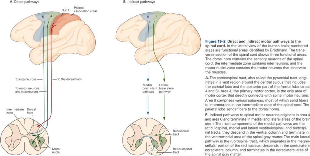
Outputs
- All three areas (MI, PM, and SMA) contribute fibers to the corticospinal/corticobulbar tracts. However, MI provides the only pathway that controls digit muscles. SMA and PM affect distal muscles through their projections to MI.
- Cerebellum, via pontine nuclei
- Basal ganglia
Functions of the primary motor cortex (motor execution)
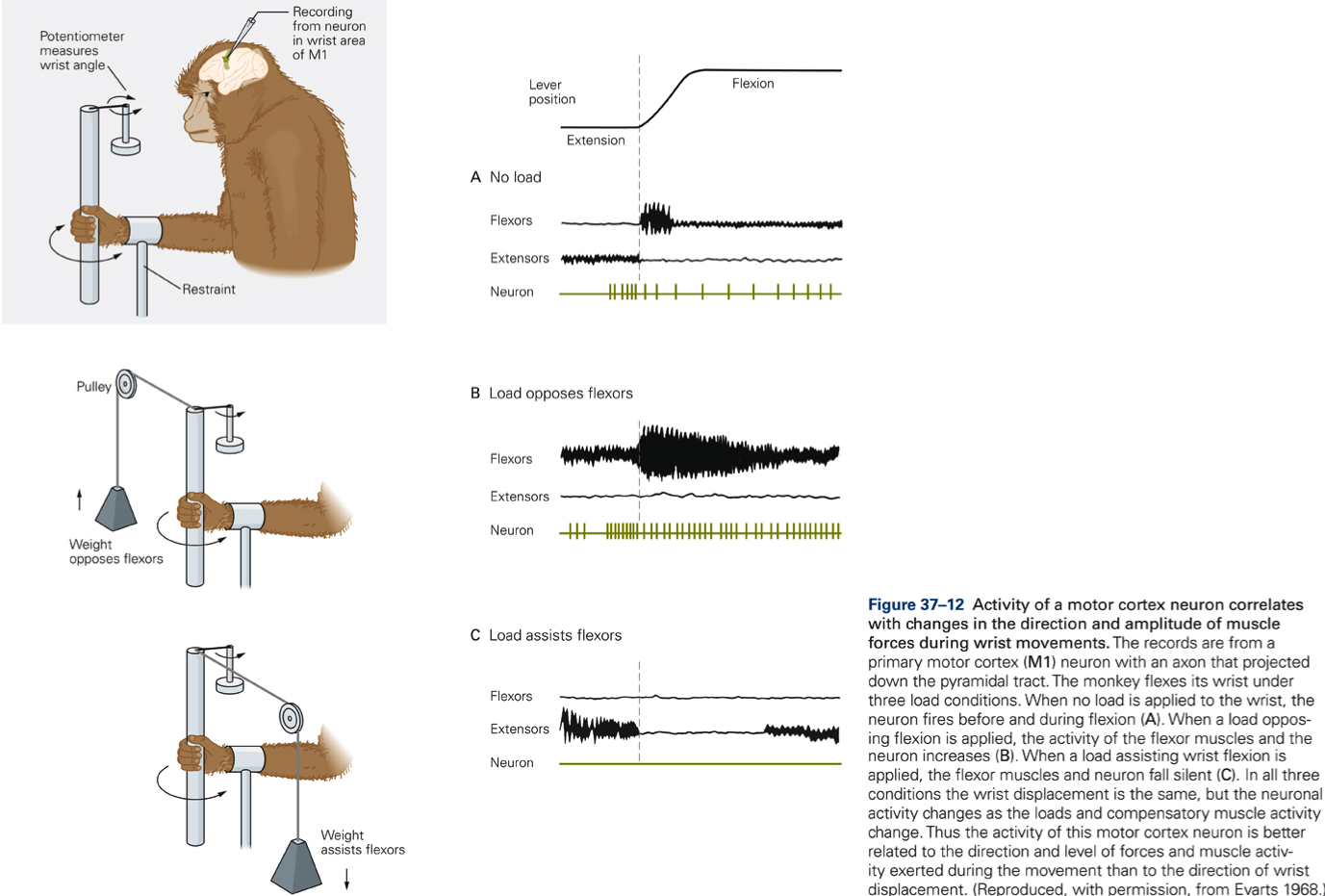
Discharge of individual neurons represent motor command signals that specify movement force
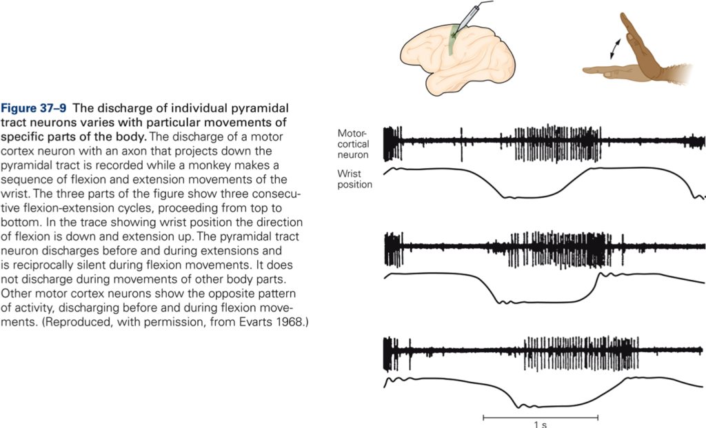
Discharge of individual neurons in primary motor cortex were recorded during same amplitude wrist movement under different loading conditions.
Key Takeaways
Evarts, 1968
- Discharge precedes movement onset (necessary for signal to be motor command).
- Different populations of neurons are active during flexion then during extension movements
- Discharge frequency codes movement force (not movement velocity, amplitude, or direction)
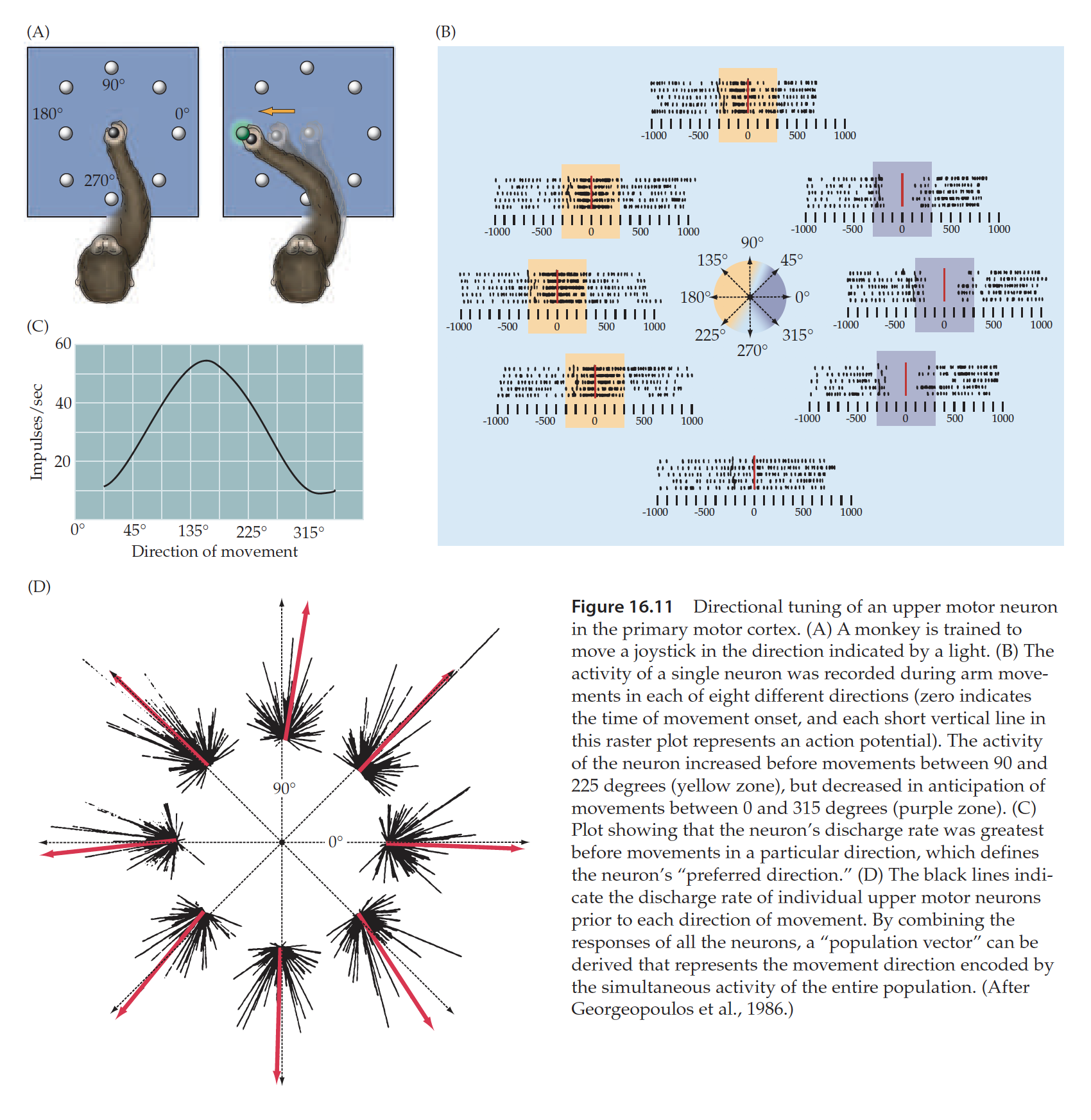
Discharge of populations of neurons specifies movement direction.
Individual neurons are broadly tuned for movement direction, i.e. they are activated optimally for one specific (preferred) direction and are activated less for directions that differ from the preferred direction.
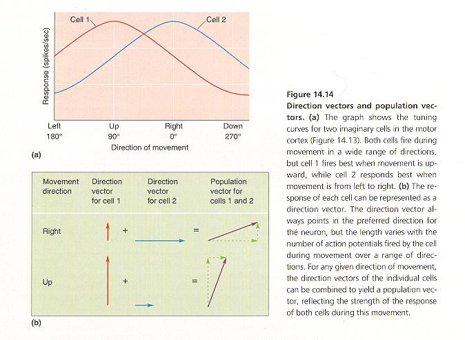
 Vector hypothesis
Discharge of an individual neuron can be represented by a vector (direction of vector points in preferred direction; length of vector is proportional to amplitude of discharge). The sum of vectors (population vector) points in (predicts) the direction of the movement.
Key Takeaways
Georgeopoulos et al. 1986
- Neurons in the primary motor cortex are broadly tuned for movement direction.
- The direction vector for an individual neuron always points in the neuron’s preferred direction.
- The population vector (sum of direction vectors of individual neurons) predicts movement direction.
