
M2 | Spinal Mechanisms for Sensorimotor Integration
Spinal reflexes
Stretch reflex
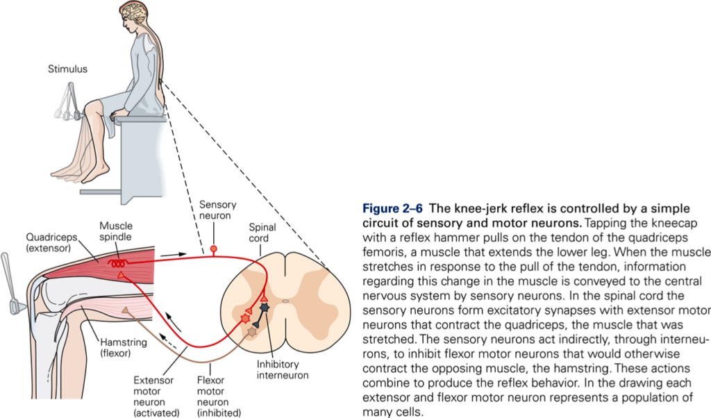
Stretch reflexes are the simplest of all spinal reflexes. An example of a stretch reflex is the knee jerk. A sharp tap on the patellar tendon stretches the quadriceps muscle, which produces extension of the knee. Thus, the stretch reflex contracts the muscle that is lengthened (homonymous muscle).
Thomas Jesell demonstrates the knee jerk, a clinical assessment tool and important component of the physical exam [Jessell, T. M. Demonstration of the knee jerk. HHMI’s BioInteractive Video Clips: Howard Hughes Medical Institute].
Spinal circuitry
The sensory neurons involved in the stretch reflex are the Ia afferents from the muscle spindle. The cell bodies of these cells are located in the dorsal root ganglion. Their axon bifurcates, one branch goes to the muscle, the other enters the spinal cord through the dorsal roots and terminates directly on all motoneurons that innervate the muscle in which the spindle is located. Because the stretch reflex only involves one synapse, it is also known as the monosynaptic reflex. The stretch reflex is a common diagnostic tool. Weak stretch reflexes indicate disorder of one or more components of the reflex circuit.
Long-loop component
When the muscle stretches in response to the tap on the tendon, information is also transmitted by afferent (sensory) neurons to higher centers of the CNS. Some neurons of the motor cortex receive input from the same muscle to which they project. Thus, motor cortex works in parallel with the spinal stretch reflex. The circuit through the motor cortex is called the long-loop component of the stretch reflex or functional stretch reflex.
Stretch reflexes regulate muscle tone
Muscle tone: force with which muscle resist being lengthened.
Functions of muscle tone:
- Maintains posture
- Allows muscles to store energy
- Smoothes movements.
Spinal integration
Key Takeaways
- Spinal circuitry is critically important for all movements (reflexes, automatic movements, and voluntary movements) because both peripheral and central motor signals must interface with spinal circuitry to access and influence muscles.
- Interneuronal connections are important for linking muscles together into functional units that provide motor coordination.
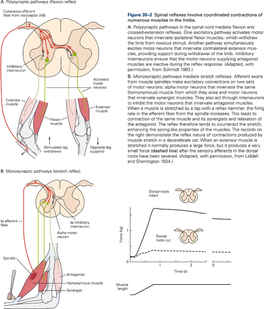
Most spinal reflexes are polysynaptic; they have one or more interneurons in between the sensory and motor neuron.
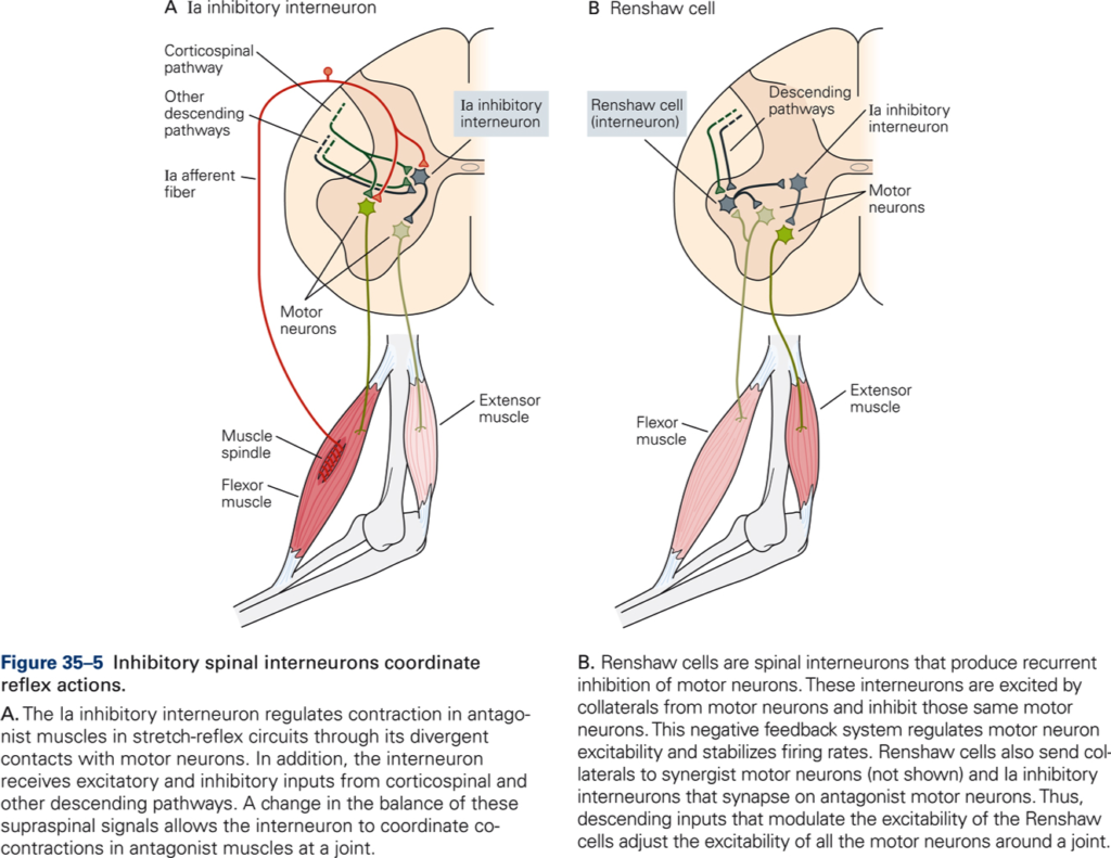
Ia inhibitory interneurons
Ia afferents also terminate on group Ia inhibitory interneurons that project to motoneurons of the antagonist (opposing) muscles. Thus, during the knee jerk, stretching of the knee extensor muscle results in activation of the knee extensor and inactivation of the knee flexor muscles, which result in movement in the direction that counteract the stretch. This pattern of connection is called reciprocal innervation; this form of coordination is not only useful for stretch reflexes but is common in many voluntary movements. Descending axons terminate directly on excitatory motoneurons and in addition terminate on Ia inhibitory inter neurons higher motor centers do not need to send separate commands to agonist and antagonist muscles, thus simplifying control.
Ia inhibitory neurons are also involved in the regulation of cocontraction (the simultaneous activation of agonist and antagonist muscles). Cocontraction is advantageous when stiffer joints are required, for example, during precision movements. Ia inhibitory neurons receive inhibitory and excitatory inputs from all major descending motor pathways by regulating balance of activity the brain can control the relative amount of joint stiffness.
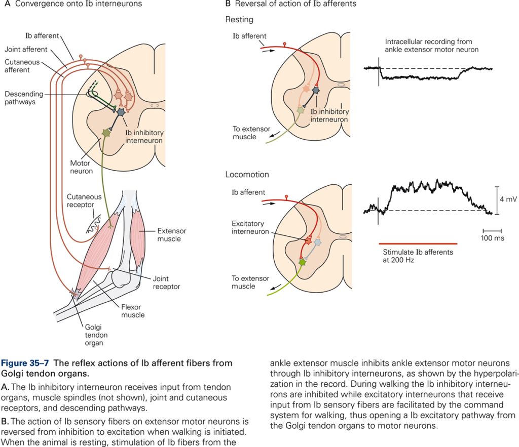
Ib inhibitory interneurons
Golgi tendon organ reflex
Negative feedback loop for regulating muscle force. In addition to GTO input, Ib inhibitory interneurons receive inputs from many sources: e.g. low threshold cutaneous, joint receptors, descending inputs.
Integration of muscle and cutaneous information
Spinal mechanism that underlie fine control of exploratory movements, such as during active touch, depend on integration of muscle and cutaneous information. Upon contact, force is immediately inhibited by activation of GTOs; cutaneous afferents soften contact. Descending signals modulate strength of inhibition, i.e. increase inhibition if the object is fragile, decrease inhibition if more forceful contact is required.
Cutaneous reflexes
Watch: Reflex Arcs – Narrated animation & Quiz
Stimulation of skin initiates complex protective and postural reflexes. Stimulation of many areas of skin causes reflex contraction of specific muscles. For example, stroking abdomen causes contraction of abdominal muscles (local sign).
Reflex effect depends on quality of stimulus:
- Lightly stroking the bottom of the foot flexion of toes/foot elicits the plantar reflex
- Light pressure on the bottom of the foot generalized extensor response in whole leg elicits the extensor trust reflex
- A painful stimulus (pin prick, pinch) applied to same area results in contraction of all flexor muscles in limb flexor withdrawal reflex. The withdrawal reflex activates ipsilateral flexors and inhibits ipsilateral extensor motoneurons. The same stimulus causes the opposite response on the contralateral side of the body: activation of extensors and inhibition of flexor motoneurons. This is called the crossed extensor reflex.
