M5 | Integrative action of the sensorimotor system – Visual system | Association cortices
Visual system
Functional organization of the visual pathways
Watch: Visual pathways in the human brain
Connectivity
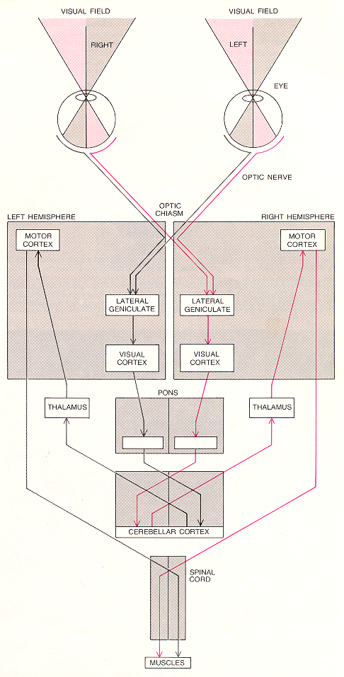
Retinal ganglion cells (RGC, output neurons of retina) project via the lateral geniculate nucleus (LGN) to the primary visual cortex (= area 17). RGC axons carrying information from half of the visual field from each eye combine at the optic chiasm. The information is transmitted via the LGN to the visual cortex in the opposite hemisphere of the brain.
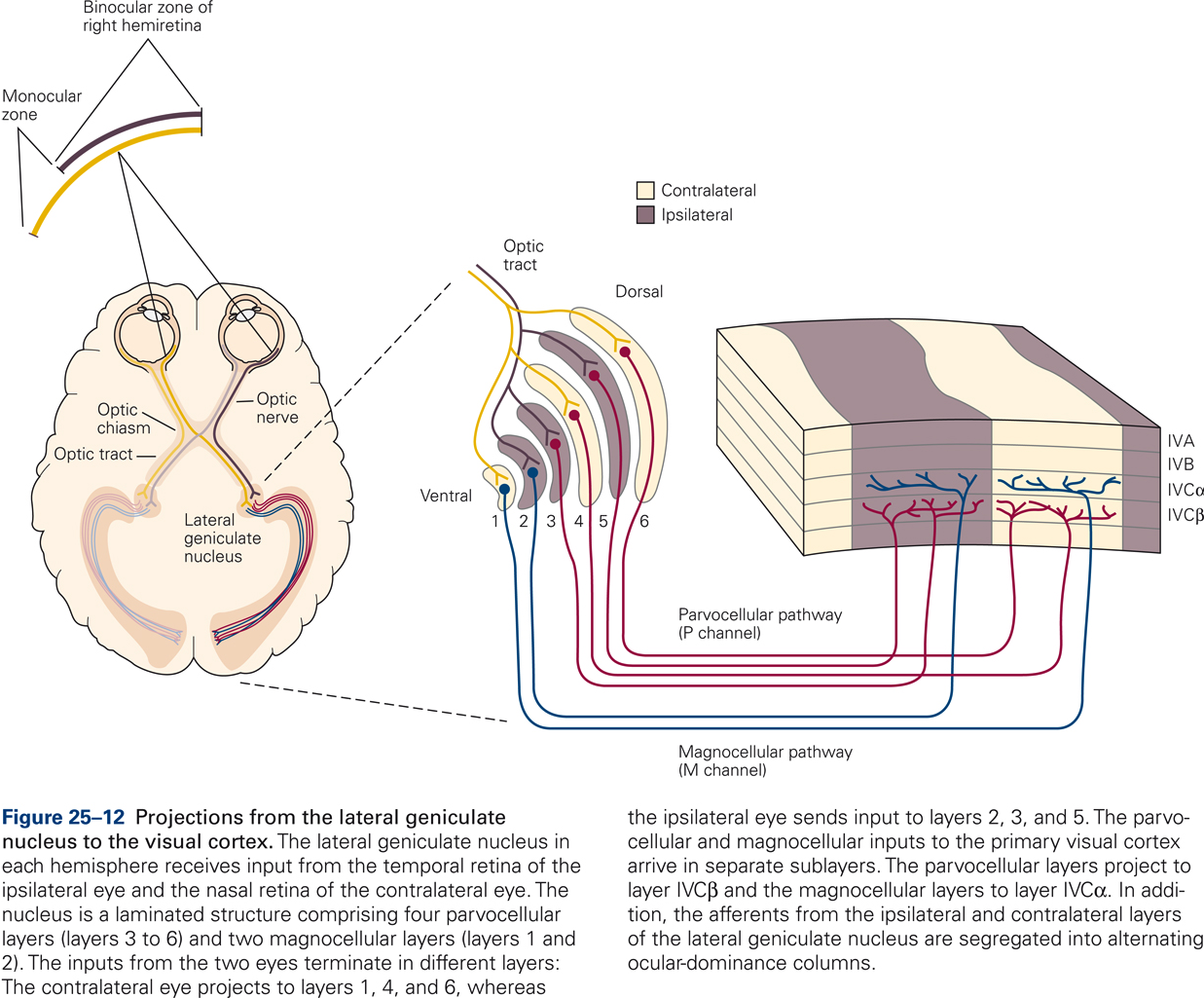
The right half of the visual field is imaged on the temporal half of the retina of the left eye and on the nasal half of the retina of the right eye. RGC output from these retinal areas is transmitted via the left LGN to the left visual cortex. Although each LGN receives information from both eyes, the information from the two eyes is first combined in the visual cortex.
Mapping is retinotopic
The retinal image of the visual world is accurately reproduced in the LGN and visual cortex; thus each of the structures has a highly detailed map of the visual world, that is, adjacent points in the visual world are processed by adjacent RGC, and LGN and visual cortical neurons.
Central parts of the visual field are overrepresented
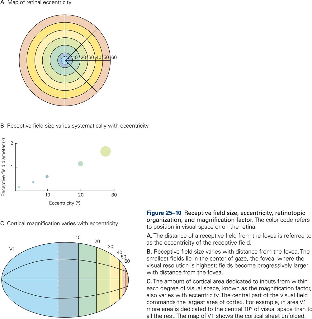
The central part of the retina (fovea) provides the highest spatial resolution of the visual world. The density of RGCs in the fovea is higher than in more peripheral regions, and the receptive fields of RGCs in the fovea are smallest. In addition, RGCs in the fovea receive inputs from photoreceptors with the highest spatial resolution (cones). As the information is passed along the visual pathways, from RGCs to LGN, to visual cortex, at successive stages more neurons are devoted to analyzing information from the central few degrees of vision (compare with the distortion in the somatotopic maps in sensorimotor cortices).
Information processing along the visual pathways
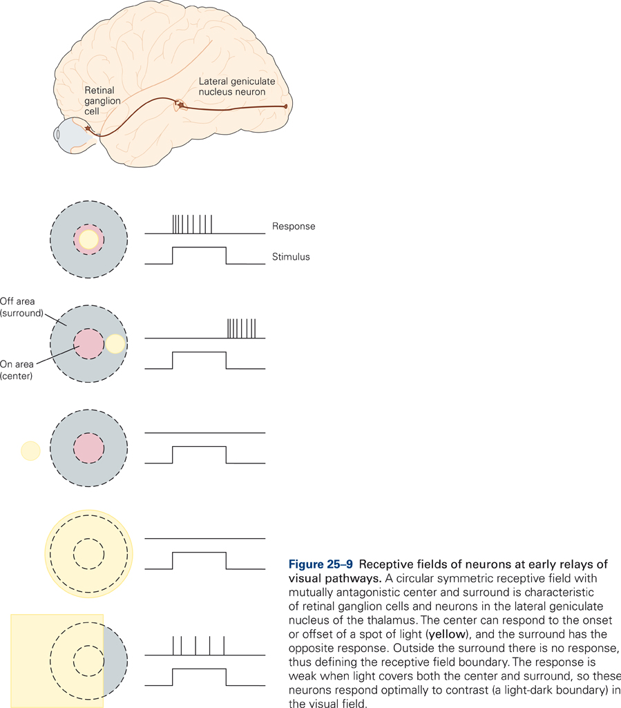
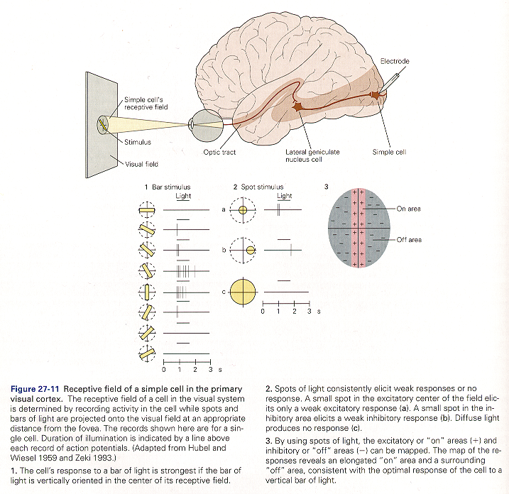
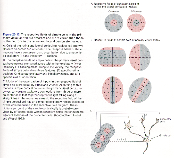
Retinal ganglion and LGN cells respond best to spots of light
RGCs and LDN cells respond optimally to contrast in their receptive fields. Their receptive fields are circular, divided into a center area and a “donut-shaped” surround. On-center cells are excited by light in the center and inhibited by light in the surround. Off-center cells have opposite response properties.
Watch: Information Processing in the Retina – Narrated animation & Quiz
Neurons in the visual cortex respond best to oriented line stimuli
David Hubel and Torsten Wiesel, working at Harvard University, won the Nobel prize (1981) for their studies in the 1960s that revealed the functional architecture of the primary visual cortex.
Media: Kaltura Mediaspace
Receptive fields of visual cortical neurons consist of excitatory and inhibitory regions that are rectangular.
Media: Kaltura Mediaspace
Higher visual areas
Media: Kaltura Mediaspace
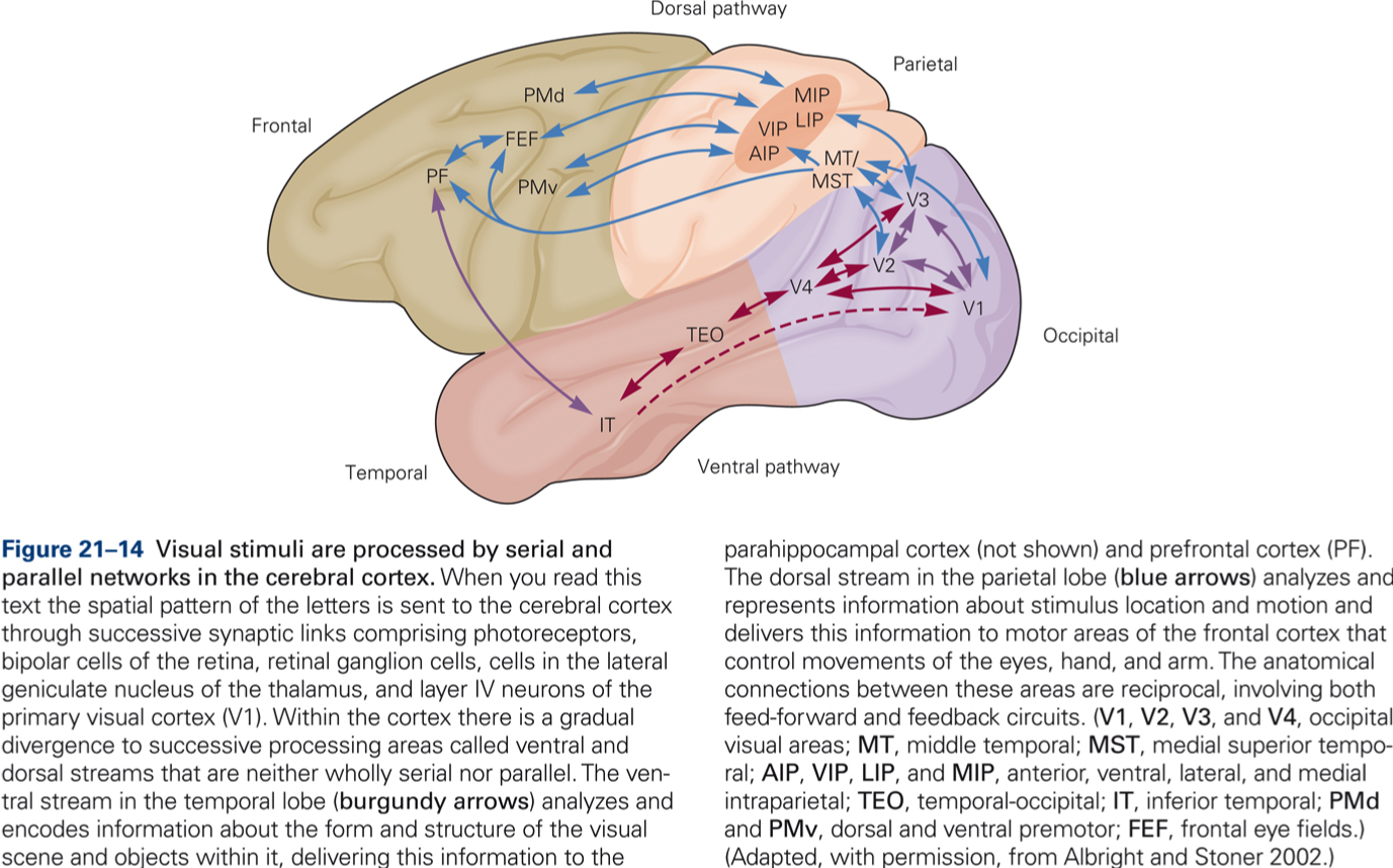
Vision for action
Parietal cortex, MT, MST: movement analysis and spatial localization.
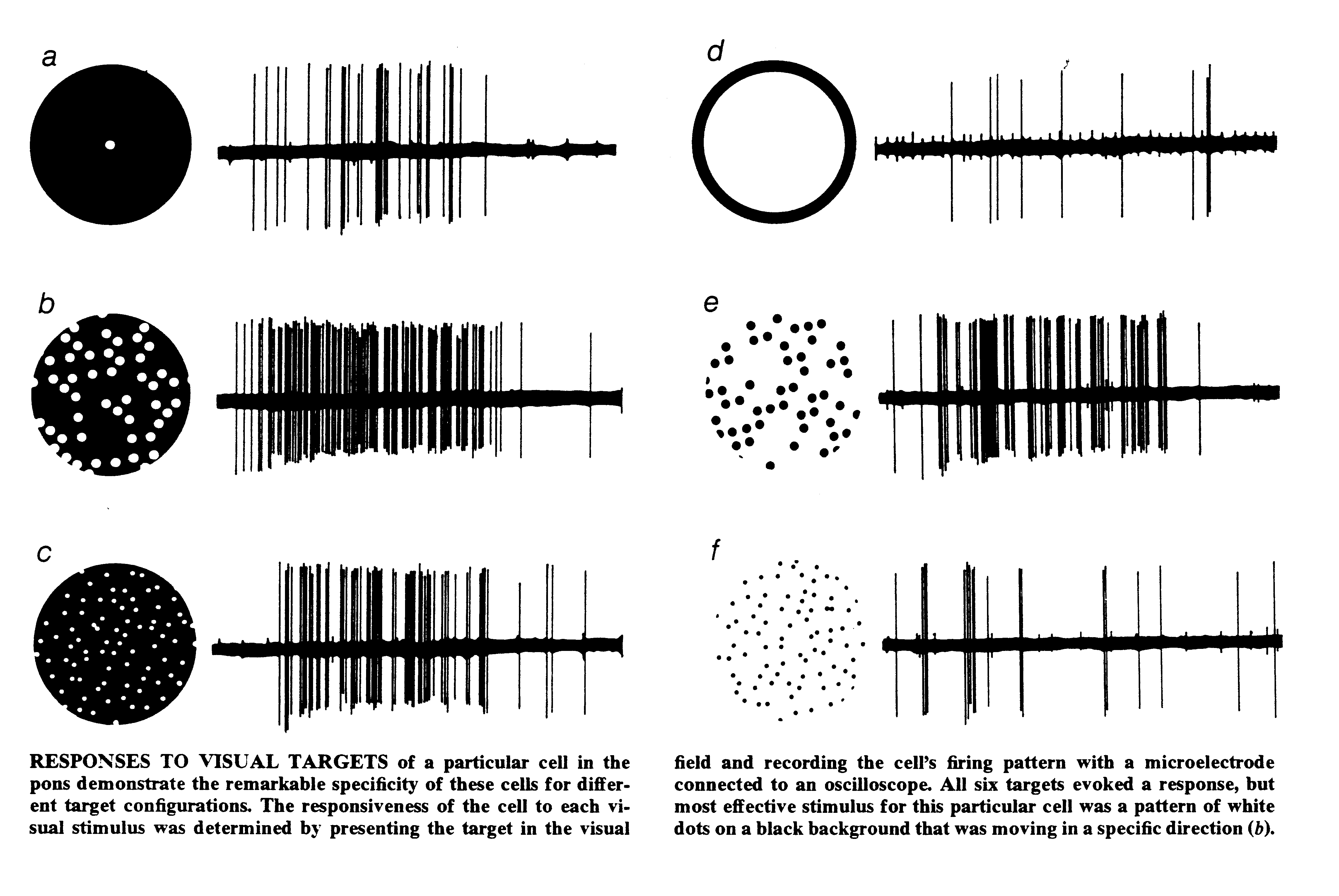
Cells in the pontine nuclei respond best to whole-field stimuli
Vision for perception
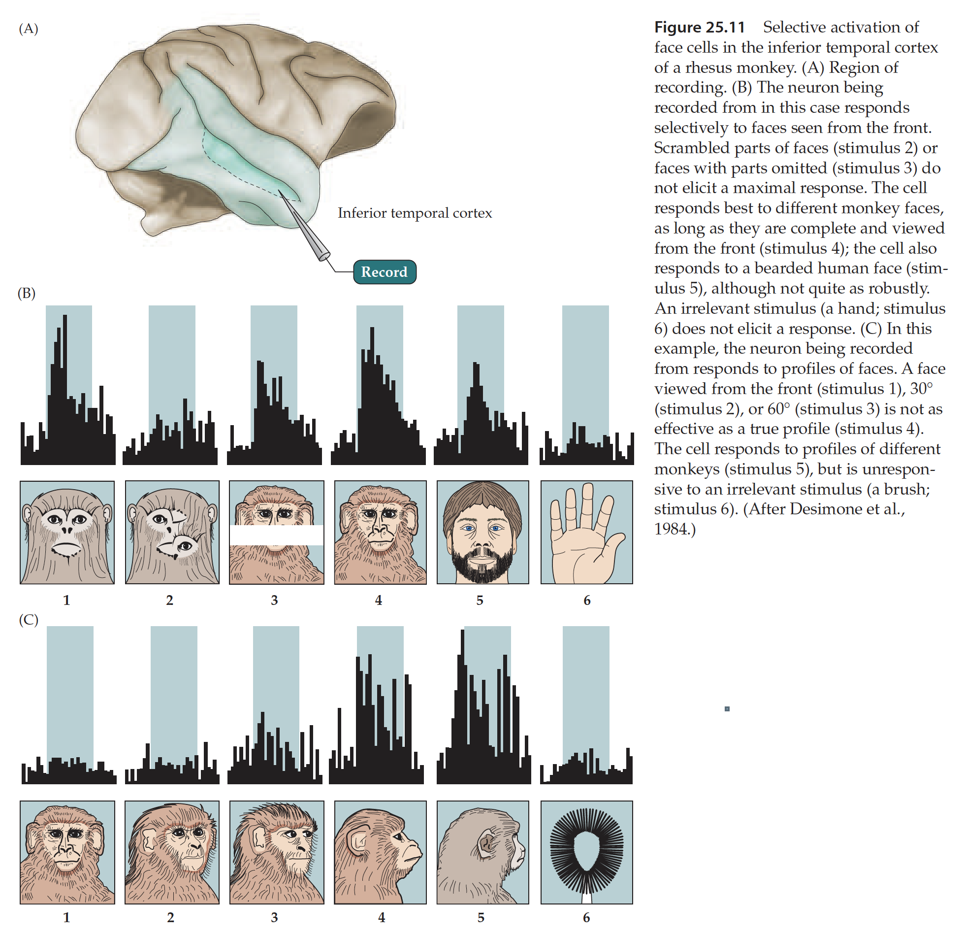
Inferior temporal cortex: face recognition and complex forms