
Module 19: The Brain Part I: Cerebrum, Diencephalon, Cerebellum, and Brainstem
Learning Objectives:
By the end of this class, students will be able to:
- Explain the orientation of the brain utilizing anatomical terms.
- Explain hemispheric lateralization.
- Describe the gray and white matter in the brain, including location, contents, and general functions.
- Describe the lobes of the brain and their components and functions.
- Discuss the various regions of the brain involved in higher-order processes, such as written and spoken language, cognition, and memory.
- Describe limbic system structures and cerebral nuclei of the brain and explain their functions.
- Explain the functions and location of the components of the diencephalon, including the endocrine functions of the hypothalamus.
- Explain the structure and function of the cerebellum.
- Explain the anatomy and functions of the three parts of the brain stem.
Terms to Know
|
Cerebrum
|
The Brain: Introduction
Diencephalon
Pituitary Gland
Cerebellum
Brainstem
*Covered only in lecture, not in this text |
The brain is made up of the cerebrum, cerebellum, and brainstem, and each region contains many smaller structures. The brain controls our thoughts, movements, emotions, senses, and so much more. You could get an entire degree learning about this amazing organ, so this module, along with the following module, will provide a brief overview of the different parts of the brain.
Introduction to the cerebrum
*This content will be covered in the assignment, primarily as self-study. Portions will be briefly reviewed in lecture.
The cerebrum makes up the largest portion of the brain. Many of the higher neurological functions, such as memory, emotion, language and consciousness, are the result of cerebral function. Beneath the cerebral cortex are sets of nuclei known as subcortical nuclei (subcortical structures) that augment cortical processes.
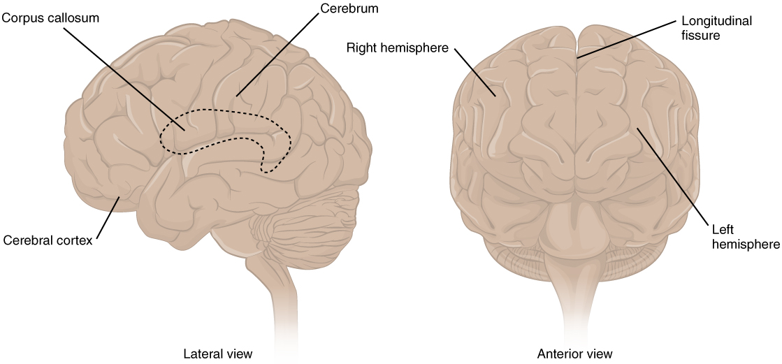
The surface of the brain contains several ridges, called gyri (singular = gyrus) and grooves between the ridges, called sulci (singular = sulcus). These folds greatly increase the surface area of the cortex, responsible for higher-order thinking. There is a large separation between the two sides of the cerebrum called the median longitudinal fissure. It separates the cerebrum into two distinct halves, a right and left cerebral hemisphere. The two hemispheres are anatomically identical but functionally different. These differences are referred to as hemispheric lateralization. For example, visuospatial processes, imagination, music, and artistic skills are generally associated with the right hemisphere. Analytical thinking and math skills are generally associated with the left hemisphere, and language centers are only located in the left hemisphere (with rare exceptions).
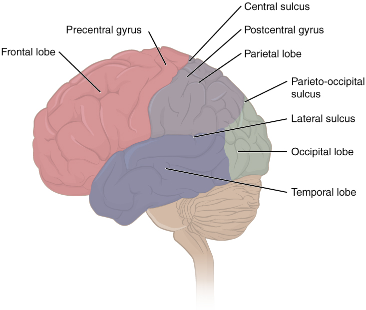
Specific sulci separate the cerebrum into four major regions, or lobes. The lateral sulcus, also known as the Sylvain fissure, separates the temporal lobe from the frontal and parietal lobes. The temporal lobe is located near the ears. The parietal lobe posteriorly and frontal lobe anteriorly, are separated from each other by the central sulcus. The posterior region of the cortex is the occipital lobe, which is separated from the parietal lobe by the the parieto-occipital sulcus on the medial aspect of each hemisphere.
Cerebral Cortex
*This content will be covered in the assignment, primarily as self-study. Portions will be briefly reviewed in lecture.
The cerebral cortex is the thin layer of gray matter on the outside of the cerebrum. It is highly folded to fit within the limited space inside the skull, creating the sulci and gyri. This thin, extensive region of gray matter is responsible for the higher functions of the nervous system. These higher functions are distributed across various regions of the cortex, and specific locations can be said to be responsible for particular functions.
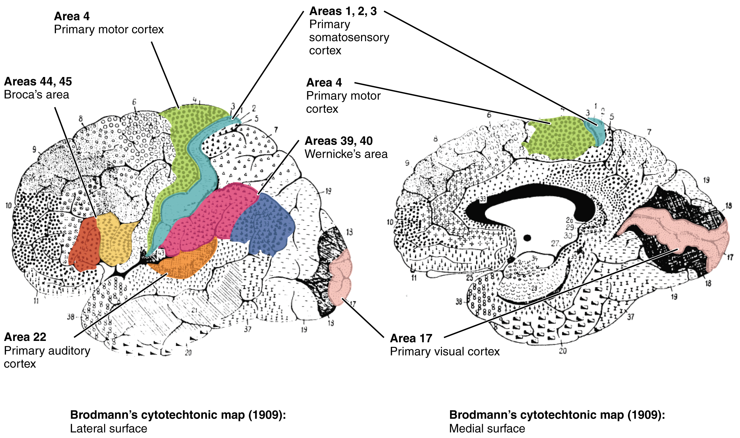
In the early 1900s, a German neuroscientist named Korbinian Brodmann performed an extensive study of the microscopic anatomy—the cytoarchitecture—of the cerebral cortex and divided the cortex into 52 separate regions on the basis of the histology of the cortex. He compared the size, shape, and number of neurons to find anatomical differences in the various regions. The differences in neurons correspond to different functional areas as well. Therefore, the structure of the neurons in a given region of the cerebral cortex governs the function of that area of the cortex. His work resulted in a system of classification known as Brodmann’s areas, which is still used today to describe the anatomical distinctions within the cortex. We will reference a few of these regions in lecture. When we mention area BA #, we are referring to Brodmann’s areas.
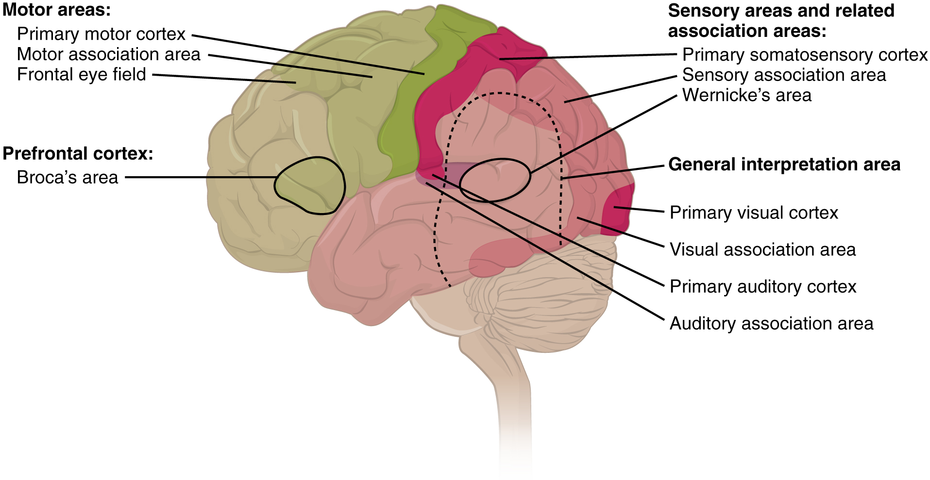
These regions can be grouped into sensory, motor, and multimodal, or integrative, areas.
- Sensory areas: process incoming sensory information
- Multimodal association areas: complex processing and integration of the different types of sensory input coming in at a given time along with memories and emotions, make a plan for how to respond
- Motor areas: execute the motor response to the sensory information
Occipital Lobe
The occipital lobe is responsible for processing visual information. We will not discuss the occipital lobe here. Instead, we will discuss visual processing in the occipital lobe with the visual system module.
Parietal Lobe
This information will be covered in lecture.
The parietal lobe is located posterior to the central sulcus and frontal lobe, anterior to the occipital lobe, and superior to the temporal lobe and lateral sulcus. This lobe of the brain is involved in sensory processing.
The primary sensory function of the parietal lobe is somatosensation, meaning the general sensations associated with the body. Posterior to the central sulcus is the postcentral gyrus which houses the the primary somatosensory cortex. This region is identified as Brodmann’s areas 3, 1, and 2. This area is in charge of the initial processing of all tactile senses, including touch, pressure, tickle, pain, itch, and vibration. Posterior regions of the parietal lobe include somatosensory association areas and sensory integration areas that integrate other senses, such as visual stimuli, as well.
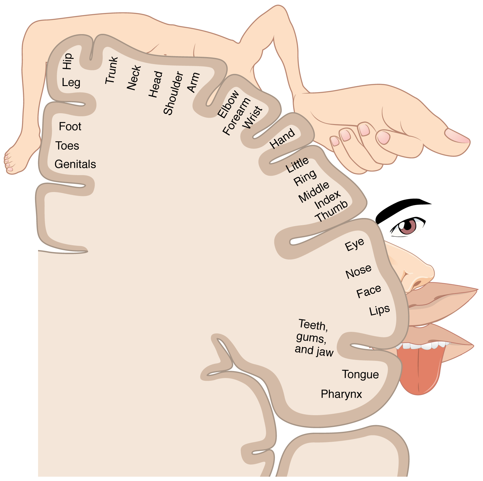
The term homunculus comes from the Latin word for “little man” and refers to a map of the human body that is laid across a portion of the cerebral cortex. Somatosensory receptors in the body are essentially mapped onto the somatosensory cortex, forming a somatosensory homunculus. In the somatosensory cortex, the external genitals, feet, and lower legs are represented on the medial surface of the gyrus within the longitudinal fissure. As the gyrus curves out of the fissure and along the surface of the parietal lobe, the body map continues through the thighs, hips, trunk, shoulders, arms, and hands. The head and face are just lateral to the fingers as the gyrus approaches the lateral sulcus. The areas of the body that are capable of fine touch sensation, such as the fingers and lower face, have a larger representation on the cortex. Less sensitive areas of the body, such as the shoulders and back, are mapped to smaller areas on the cortex.
Frontal Lobe
This information will be covered in lecture.
Anterior to the central sulcus is the frontal lobe. The frontal lobe is involved in motor activity a well as higher-order thinking and personality.
The primary motor cortex is located in the precentral gyrus of the frontal lobe, just anterior to the central sulcus. Neurons originating in this cortical region are the upper motor neurons that travel to the spinal cord to synapse with lower motor neurons that eventually innervate skeletal muscles.
The primary motor cortex is arranged in a similar fashion to the primary somatosensory cortex, in that it has a topographical map of the body, creating a motor homunculus. The neurons responsible for musculature in the feet and lower legs are in the medial wall of the precentral gyrus, with the thighs, trunk, and shoulder at the crest of the longitudinal fissure. The hand and face are in the lateral face of the gyrus. Also, the greatest amount of cortical space is given to muscles that perform fine, agile movements, such as the muscles of the fingers and the lower face. The “power muscles” that perform coarser movements, such as the buttock and back muscles, occupy much less space on the motor cortex.
Two important regions that assist in planning and coordinating movements are located adjacent to the primary motor cortex. The premotor cortex aids in controlling movements of the core muscles to maintain posture during movement, whereas the supplemental motor area is hypothesized to be responsible for planning and coordinating movement. The supplemental motor area also manages sequential movements that are based on prior experience (that is, learned movements). Neurons in these areas are most active leading up to the initiation of movement.
The prefrontal cortex, the anterior portion of the frontal lobe, is responsible for higher-cognitive functions like planning, making decisions, judgement, multitasking, and more. These functions are called executive functions, as many lead to goal-directed behavior. The functions of the prefrontal cortex are integral to the personality of an individual, because it is largely responsible for what a person intends to do and how they accomplish those plans. The prefrontal lobe is responsible for aspects of attention, such as inhibiting distracting thoughts and actions so that a person can focus on a goal and direct behavior toward achieving that goal.
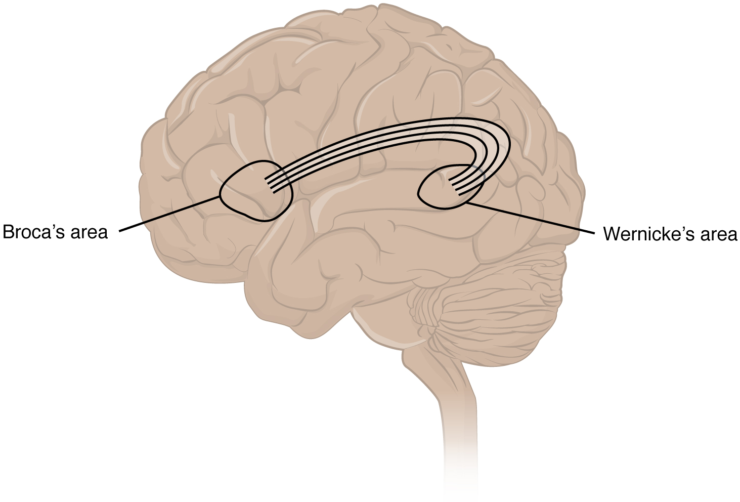
In the lateral aspect of the frontal lobe, just anterior to the region of the motor cortex associated with the head and neck, is Broca’s area. This area is responsible for controlling movements of the structures of speech production. It is only located on the left side, as only the left side of the brain is involved in language.
Another speech area is located in the temporal lobe, which you will read about next. It is called Wernicke’s area, and it is critical for the comprehension of language. Like Broca’s area, it is only located in the left hemisphere. Wernicke’s area and Broca’s area are highly connected by white matter pathways.
Temporal Lobe
This information will be covered in lecture.
The temporal lobe is located on the lateral aspect of the brain, near the ears. It contains three parallel horizontal gyri on its lateral aspect, called the superior, middle, and inferior temporal gyri. One function of the temporal lobe is audition, or hearing. The primary auditory cortices, Brodmann’s areas 41 & 42, are in the superior temporal lobe. This is the first cortical area involved in processing hearing.
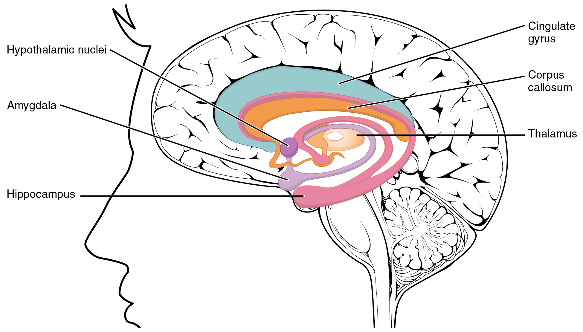
The limbic system is a collection of structures involved in emotion, memory, and behavior. While not all structures of the limbic system are located in the temporal lobe, two key structures are: the amygdala and hippocampus.
Amygdala
The amygdala is located in the medial temporal lobe. It is critical for processing emotions and tying emotions to our sensations and our current environment, especially fear. For example, when you are anxious or scared, the amygdala will send signals to other parts of the brain to eventually trigger a fight-or-flight response. It is also important for regulating those emotions, along with part of the prefrontal cortex. It is highly connected to the hippocampus, which is critical for forming memories. Through that connection the amygdala plays a role in tying emotions to experiences and to memories. If a current experience reminds you of a situation that brought you fear, the amygdala will tie the fear from that memory to the current situation. In a new experience, the amygdala will work with the hippocampus to tie the emotion of that experience to the new memory.
Hippocampus
The hippocampus is located just posterior to the amygdala in the medial temporal lobe. This structure is critical for the storage of new episodic memories into long-term memory. These memories include those of events or experiences. This is different than working memory, in which we take in information and use it right away (located in the prefrontal cortex) and procedural memory, which includes memory for motor activities, like swinging a baseball bat (controlled by the cerebellum).
Basal Ganglia (Cerebral Nuclei)
*This content will be covered in the assignment, primarily as self-study. Portions will be briefly reviewed in lecture.
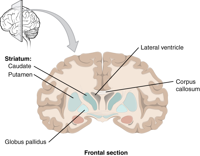
The basal ganglia (cerebral nuclei) are a set of nuclei deep in the cerebrum involved in controlling aspects of movement, including the impulse to move. The major structures of the basal nuclei that control movement are the caudate, putamen, and globus pallidus. The caudate is a long nucleus that follows the basic C-shape of the cerebrum from the frontal lobe, through the parietal and occipital lobes, into the temporal lobe. The putamen is mostly deep in the anterior regions of the frontal and parietal lobes. The globus pallidus is a layered nucleus that lies just medial to the putamen. The basal nuclei are connected with a few more nuclei in the brain stem and diencephalon that together act as a functional group that forms a motor pathway. Through these connections they assist with activities such as controlling how our arms and legs are coordinated when we walk, planning and modulating movements, and maintaining smoothness of movement. They also play a role in reward processing, motivation, and decision-making and are thought to be involved in addiction.
The Diencephalon
*This content will be covered in the assignment, primarily as self-study. Portions will be briefly reviewed in lecture.
The diencephalon consists of every structure with “thalamus” in the name: the thalamus, hypothalamus, subthalamus, and epithalamus. We won’t talk about the subthalamus in this course, and we will cover part of the epithalamus with the visual system module.
Thalamus
*This content will be covered in the assignment, primarily as self-study. Portions will be briefly reviewed in lecture.
The thalamus is a collection of nuclei that relay information between the cerebral cortex and the spinal cord or brain stem. The thalamus does not just pass the information on, it also processes that information. For example, the portion of the thalamus that receives visual information will influence what visual stimuli are important, or what receives attention. The cerebrum also sends information down to the thalamus, which usually communicates motor commands. This involves interactions with the cerebellum and other nuclei in the brain stem and basal ganglia.
There is a left and right thalamus, and the two thalami together form the walls of the third ventricle, which you will learn about in the next module.
Hypothalamus
*The general content on the hypothalamus will be covered in the assignment, primarily as self-study. Portions on the pituitary gland will be covered in lecture.
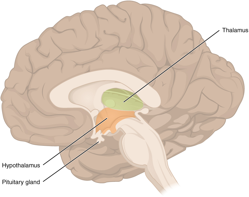
Inferior and slightly anterior to the thalamus is the hypothalamus, the other major region of the diencephalon. The hypothalamus is the executive region in charge of the autonomic nervous system and the endocrine system through its regulation of the pituitary gland. It has both neural and endocrine functions, producing and secreting many hormones. Other parts of the hypothalamus are involved in memory and emotion as part of the limbic system.
Autonomic control is based on the balance between the two divisions of the autonomic system. Coordinating that balance requires integration that begins with forebrain structures like the hypothalamus and continues into the brain stem and spinal cord.
The hypothalamus–pituitary complex can be thought of as the “command center” of the endocrine system. This complex secretes several hormones that directly produce responses in target tissues, as well as hormones that regulate the synthesis and secretion of hormones of other glands.
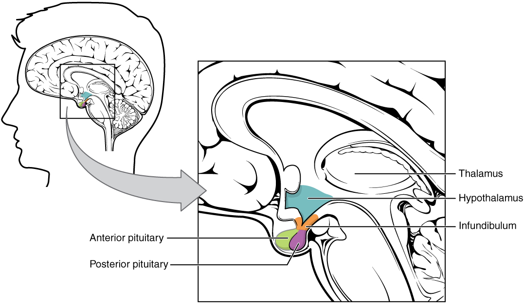
The pituitary gland consists of two lobes: the posterior pituitary (neurohypophysis) is neural tissue, whereas the anterior pituitary (also known as the adenohypophysis) is glandular tissue. The infundibulum is a “stalk” that connects the hypothalamus to the pituitary gland.
Anterior Pituitary
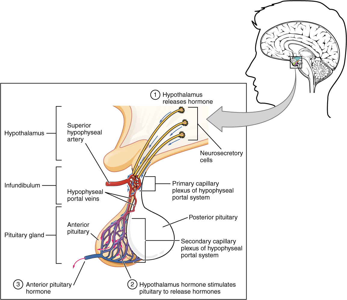
The secretion of hormones from the anterior pituitary, or adenohypophysis, is regulated by two classes of hormones. These hormones—secreted by the hypothalamus—are the releasing hormones that stimulate the secretion of hormones from the anterior pituitary and the inhibiting hormones that inhibit secretion.
Hypothalamic hormones enter the anterior pituitary through blood vessels. Within the infundibulum is a bridge of capillaries that connects the hypothalamus to the anterior pituitary. This network, called the hypothalamic-hypophyseal portal system, allows hypothalamic hormones to be transported to the anterior pituitary without first entering the systemic circulation. Hypothalamic releasing and inhibiting hormones travel through a primary capillary plexus to the portal veins, which carry them into the anterior pituitary. Hormones produced by the anterior pituitary (in response to releasing hormones) enter a secondary capillary plexus, and from there drain into the circulation.
Posterior Pituitary
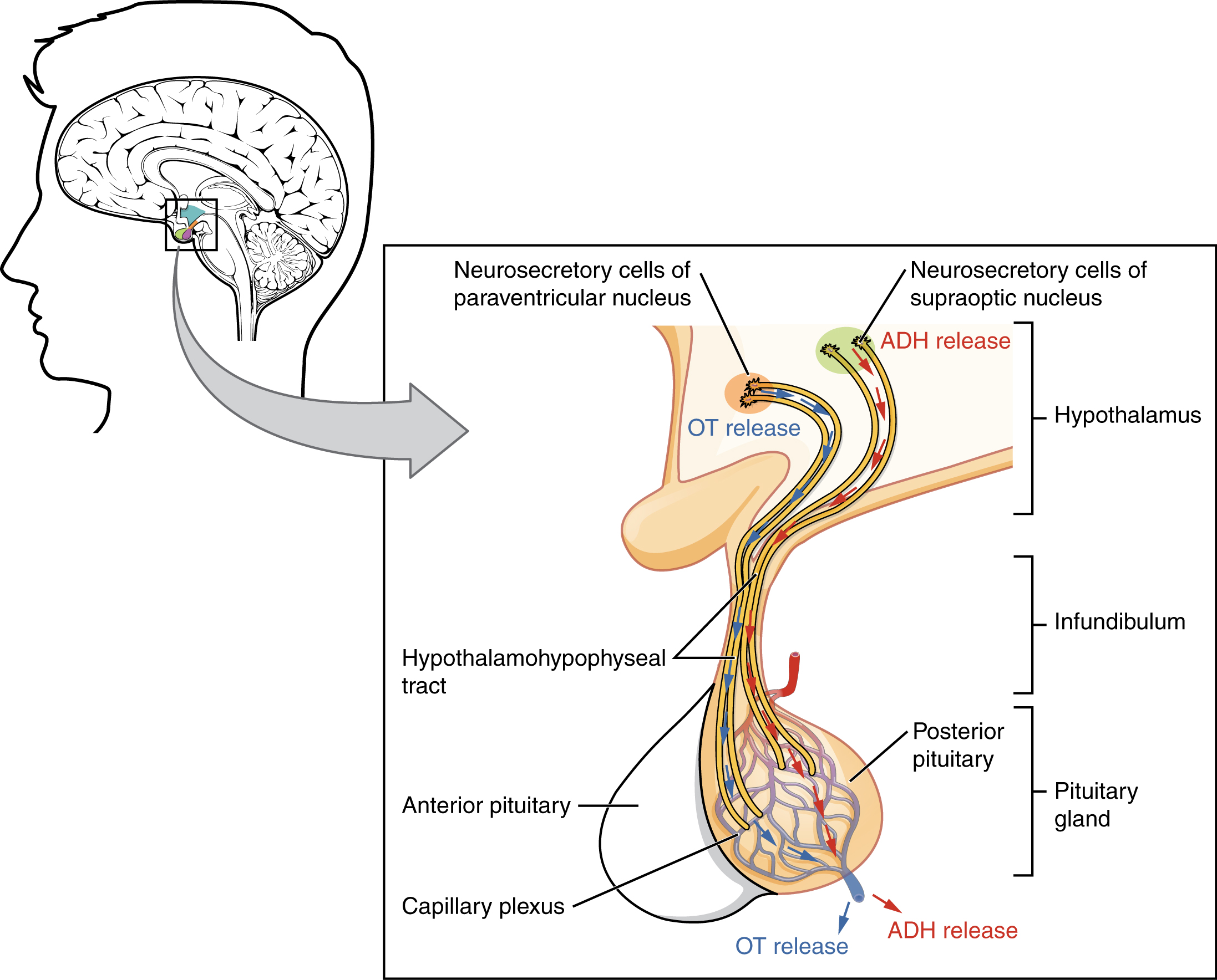
The posterior pituitary, or neurohypophysis, is an extension of the neurons of the hypothalamus. The cell bodies of these regions rest in the hypothalamus, but their axons descend as the hypothalamic–hypophyseal tract within the infundibulum, and end in axon terminals that comprise the posterior pituitary. The posterior pituitary gland does not produce hormones, but rather stores and secretes hormones produced by the hypothalamus. In response to signals from the same hypothalamic neurons, the hormones are released from the axon terminals into the bloodstream.
The Cerebellum
*This content will be covered in the assignment, primarily as self-study. Portions will be briefly reviewed in lecture.
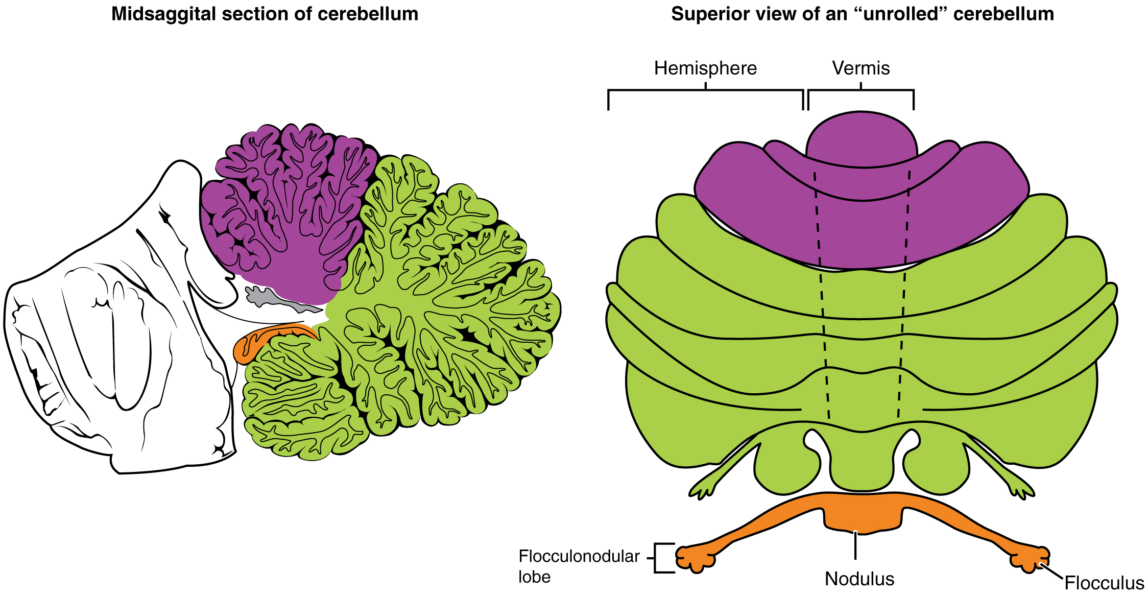
The cerebellum, as the name suggests, is the “little brain,” and it accounts for approximately 10 percent of the mass of the brain. It is covered in gyri and sulci like the cerebrum, though they appear more feathered or leaf-like, which is why they are called the folia. The cerebellum is largely responsible for comparing information from the cerebrum with sensory feedback from the periphery through the spinal cord. It is the master coordinator, coordinating primarily motor control but also higher cognitive functions and emotions.
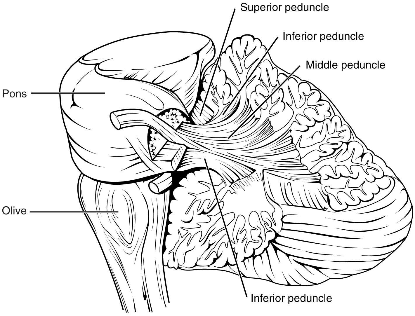
The cerebellum has an anterior and posterior lobe, forming the majority of the structure. These lobes are separated by the primary fissure. It also contains a smaller flocculonodular lobe anteriorly. The vermis is the midline portion of the cerebellum between the two cerebellar hemispheres. The cerebellum has connections to portions of the brainstem, spinal cord, and cerebrum. The superior, middle, and inferior cerebellar peduncles contain white matter pathways travelling to or from the cerebellum. The peduncles directly connect the cerebellum to parts of the brainstem, but the pathways will continue to or from these other regions of the CNS.
Sensory information from the periphery travels to the cerebellum and are compared with the descending commands from the cerebrum. If the primary motor cortex of the frontal lobe sends a command down to the spinal cord to initiate walking, a copy of that instruction is sent to the cerebellum. Sensory feedback from the muscles and joints, proprioceptive information about the movements of walking, and sensations of balance are sent to the cerebellum, and the cerebellum compares them. If walking is not coordinated, perhaps because the ground is uneven or a strong wind is blowing, then the cerebellum sends out a corrective command to compensate for the difference between the original cortical command and the sensory feedback.
Brain Stem
This information will be covered in lecture.
The brainstem consists of the midbrain superiorly, pons in the middle, and medulla oblongata inferiorly. Collectively they are responsible for many unconscious functions. It is also the origin for ten of the twelve cranial nerves. The major ascending and descending pathways between the spinal cord and brain, specifically the cerebrum, pass through the brain stem. A diffuse region of gray matter throughout the brain stem, known as the reticular formation, is related to sleep and wakefulness, such as general brain activity and attention.
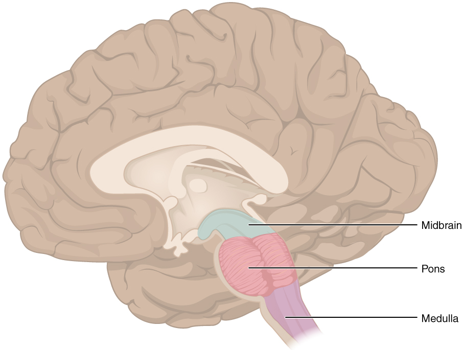
Midbrain
The midbrain is a small region between the thalamus and pons. The cerebral aqueduct passes through the center of the midbrain. The cerebellar peduncles connect the midbrain to the cerebellum.
The tectum is composed of four bumps known as the colliculi (singular = colliculus). Together they are called the corpora quadrigemina. The inferior colliculus is the inferior pair of these enlargements and is important for auditory reflexes. The superior colliculus is the superior pair and is important for visual reflexes.
The midbrain also contains two structures involved in regulating motor activity. The red nucleus is involved in motor regulation and muscle tone through connections with other parts of the brain. The substantia nigra is part of a neural circuit with the basal ganglia, contributing to motor control and modulation.
Pons
The pons is visible on the anterior surface of the brain stem as the thick belly attached to the cerebellum. The pons is the main connection between the cerebellum and the brain stem, particularly through the middle cerebellar peduncle. Gray matter in the pons contains neurons receiving descending input from the forebrain that is sent to the cerebellum as well as the pontine respiratory center.
Medulla
The medulla is the most inferior portion of the brainstem. It contains a significant amount of white matter, which is continuous with the white matter of the spinal cord. The pyramids are two vertical ridges on the anterior aspect of the medulla formed by the axons of the corticospinal tract. The pyramidal decussation is the area on the inferior aspect of the anterior medulla where the corticospinal tract fibers cross. It appears as a small area where the groove between the two pyramids briefly disappears.
The medulla contains nuclei referred to as the cardiovascular center, which controls the smooth and cardiac muscle of the cardiovascular system through autonomic connections. The medulla also contains a medullary respiratory center that communicates with the respiratory center of the pons to regulate breathing.
