
Module 20: Brain II – Blood Supply, Meninges, Ventricles, and CSF
Learning Objectives:
By the end of this class, students will be able to:
- List the names, locations, and functions of the meninges of the brain.
- List the names of the ventricles in the brain.
- Describe the production and circulation of cerebrospinal fluid.
- Explain the Circle of Willis and blood flow through the brain.
Terms to Know
|
Blood Supply to the Brain
Meninges
|
Ventricles
Other
*Covered only in lecture, not in this text ^Covered in the self-study slides on Canvas |
Arterial Supply to the Brain
This information will be covered in lecture.
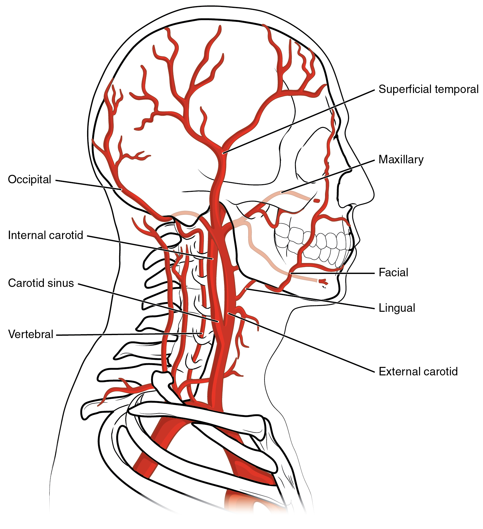
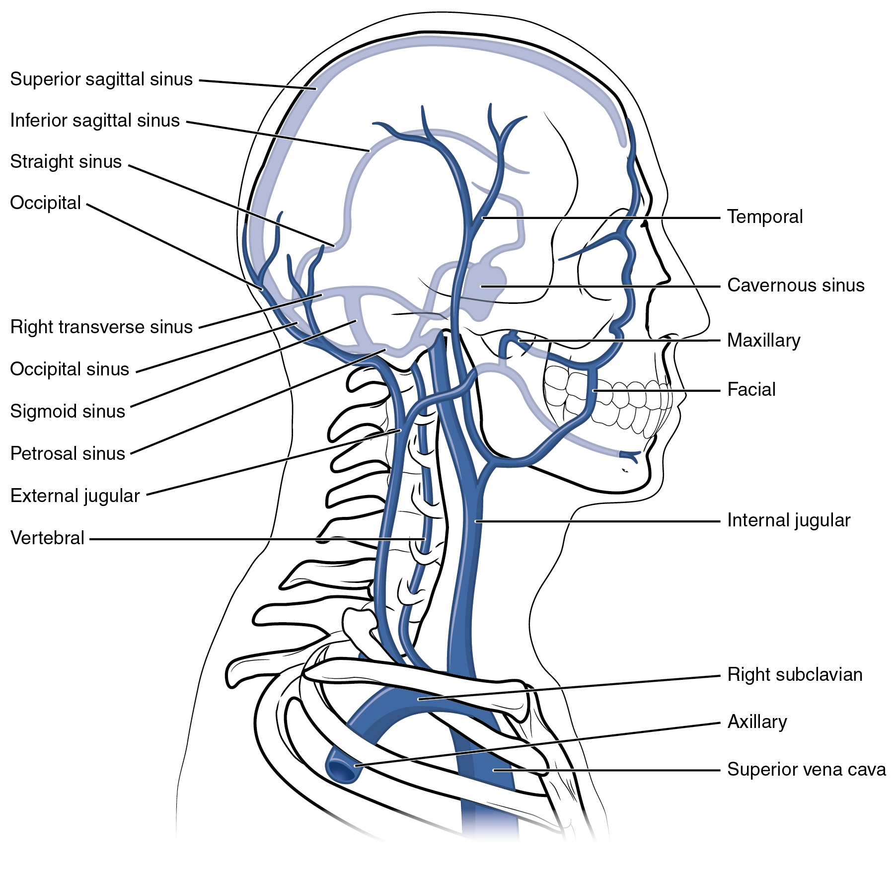
There are multiple routes for blood to get to the brain. The right and left internal carotid arteries branch from the common carotid artery on each side and enter the cranium through the carotid canal in the temporal bone. The vertebral arteries branch off of the subclavian arteries on each side, and they are protected as they pass through the neck region by the transverse foramina of the cervical vertebrae. The vertebral arteries enter the cranium through the foramen magnum of the occipital bone.
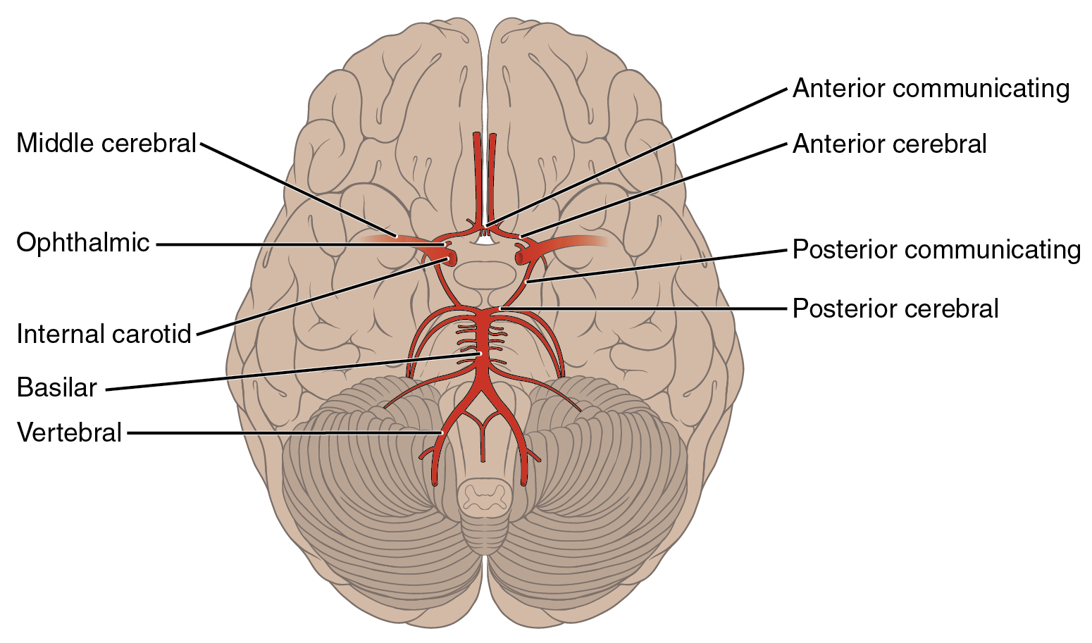
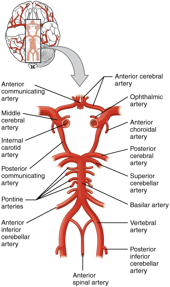
The circle of Willis is a confluence of arteries that can maintain perfusion of the brain even if narrowing or a blockage limits flow through one part. Given the central role and vital importance of the brain to life, it is critical that blood supply to this organ remains uninterrupted.
The two vertebral arteries merge into the basilar artery, which gives rise to branches to the brain stem and cerebellum. The basilar artery then ends by dividing into the right and left posterior cerebral arteries, which supply the occipital lobe and inferior aspect of the posterior temporal lobe.
The internal carotid artery continues through the carotid canal of the temporal bone and enters the base of the brain through the carotid foramen. Then it branches into the anterior and middle cerebral arteries. The anterior cerebral artery that supplies blood to the anterior, medial, and very superior portions of the frontal lobe. The middle cerebral artery supplies blood to the posterior frontal lobe and most of the temporal and parietal lobes.
The right and left anterior cerebral arteries join together to form an anastomosis called the anterior communicating artery. The initial segments of the anterior cerebral arteries and the anterior communicating artery form the anterior portion of the arterial circle. The posterior portion of the arterial circle is formed by a left and a right posterior communicating artery that runs from the internal carotid artery to the posterior cerebral artery on each side.
Venous Return from the Brain
This information will be covered in lecture.
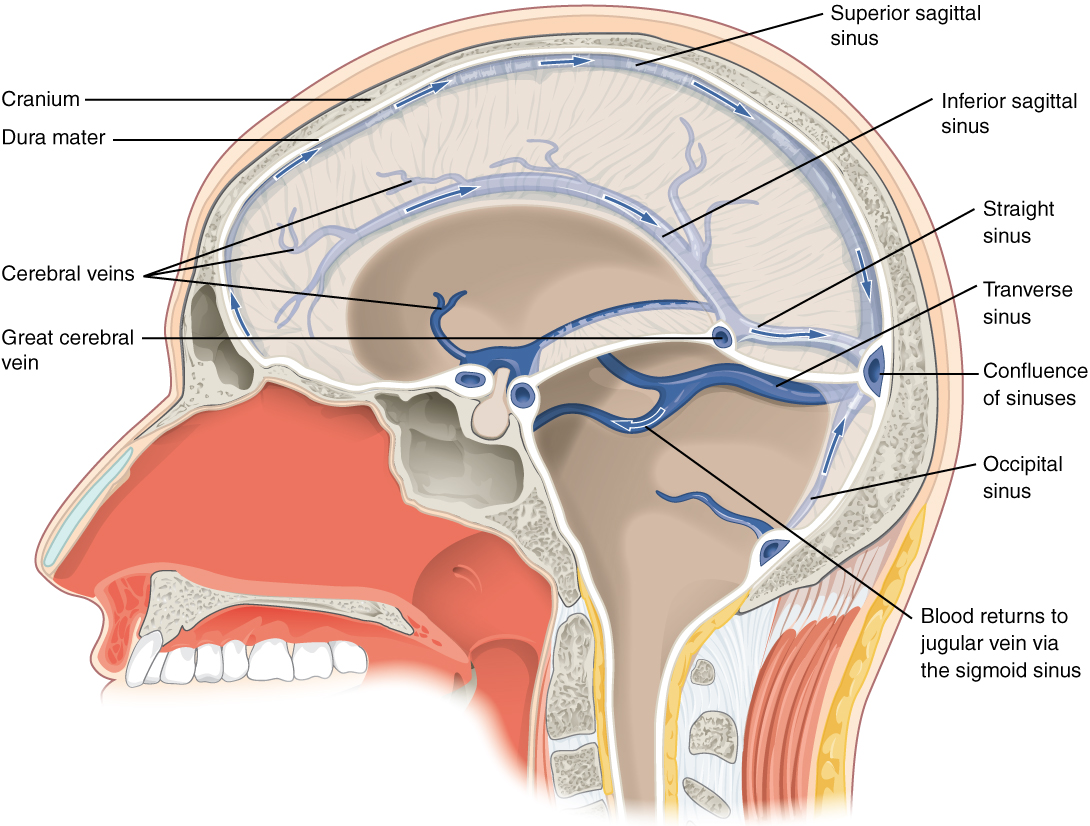
After supplying the brain with oxygen and nutrients, venules and veins merge and eventually send blood back toward the heart through a series of dural venous sinuses and veins. The superior sagittal sinus runs in the groove of the median longitudinal fissure, where it absorbs CSF from the meninges. The inferior sagittal sinus is in the inferior aspect of the falx cerebri (see the next section) within the median longitudinal fissure. It drains into the straight sinus, which joins the superior sagittal sinus to drain into the confluence of sinuses and then the transverse sinuses. The transverse sinuses connect to the sigmoid sinuses, which then connect to the jugular veins. From there, the blood continues toward the heart to be pumped to the lungs for reoxygenation.
THE MENINGES
This information is covered in the assignment. It is primarily self-study using the information here and the slides posted in the “Reading and Assignments” section of the module page on Canvas. This information will only be briefly reviewed in lecture.
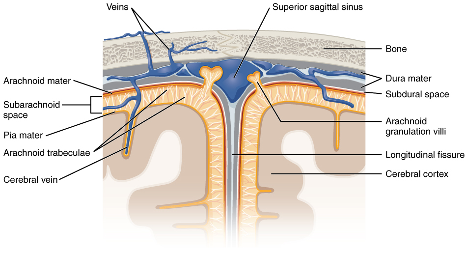
The outer surface of the CNS is covered by a series of connective tissue membranes called the meninges, which protect the brain. The dura mater is the thick outer covering, the arachnoid mater is the thin middle layer, and the pia mater is a very thin inner layer directly attached to the surface of the brain and spinal cord.
Dura Mater
Like a thick cap covering the brain, the dura mater is a tough outer covering. It encloses the entire CNS and the major blood vessels that enter the cranium and vertebral cavity. The periosteal layer is directly attached to the inner surface of the bones of the cranium. The meningeal layer is adjacent to the middle layer of the meninges.
Dural reflections are infoldings of the dura that fit into large crevasses of the brain. Two infoldings go through the midline separations of the cerebrum and cerebellum. The falx cerebri is located within the median longitudinal fissure, while the falx cerebelli is located between the right and left hemispheres of the cerebellum. The tentorium cerebelli forms a shelf-like structure between the occipital lobes of the cerebrum and the cerebellum. The dura also surrounds and supports the venous sinuses.
Arachnoid Mater
The arachnoid mater is the middle layer of the meninges. Beneath the arachnoid is a thin, filamentous mesh called the arachnoid trabeculae, which looks like a spider web, giving this layer its name. The trabeculae are found in the subarachnoid space, which is filled with circulating cerebrospinal fluid (CSF). The arachnoid emerges into the dural sinuses as the arachnoid granulations, where the CSF is filtered and drained back into the blood stream.
Pia Mater
The outer surface of the CNS is covered in the thin fibrous membrane of the pia mater. The pia extends into every convolution of the CNS, lining the inside of the sulci in the cerebral and cerebellar cortices. It also helps to support large cerebral blood vessels on the surface of the brain.
The Ventricles & Cerebrospinal Fluid
This information is covered in the assignment. It is primarily self-study using the information here and the slides posted in the “Reading and Assignments” section of the module page on Canvas. This information will only be briefly reviewed in lecture.
Cerebrospinal fluid (CSF) circulates throughout and around the CNS. CSF provides cushioning for the brain and regulates the environment around the brain. The ventricles are the open spaces within the brain where CSF circulates. CSF is produced in the ventricles by the filtering of blood in a specialized structure called the choroid plexus. Ependymal cells (a glial cell) surround blood capillaries in the choroid plexus and filter the blood to make CSF. The fluid is a clear solution with a limited amount of the constituents of blood. It is essentially water, small molecules, and electrolytes. Oxygen and carbon dioxide are dissolved into the CSF, as they are in blood, and can diffuse between the fluid and the nervous tissue. The CSF circulates through all of the ventricles to eventually emerge into the subarachnoid space where it will be reabsorbed into the blood via the arachnoid granulations.
There are four ventricles within the brain. The first two are named the lateral ventricles and are deep within each hemisphere. They are separated from each other by a thin two-layered membrane called the septum pellucidum. These ventricles are connected to the central third ventricle by the interventricular foramina (of Monro). The third ventricle is the space between the left and right diencephalon, which opens into the cerebral aqueduct that passes through the midbrain. The cerebral aqueduct opens into the fourth ventricle, which is the space between the cerebellum and the pons and upper medulla. The ventricular system opens up to the subarachnoid space from the fourth ventricle. The single median aperture and the pair of lateral apertures connect to the subarachnoid space so that CSF can leave the ventricles.
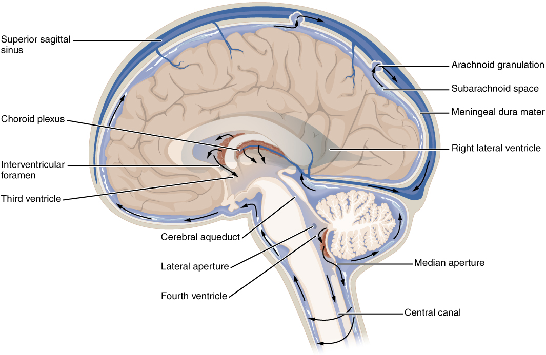
The choroid plexuses are found in all four ventricles. From the lateral ventricles, the CSF flows into the third ventricle, where more CSF is produced, and then through the cerebral aqueduct into the fourth ventricle where even more CSF is produced. A very small amount of CSF is filtered at any one of the plexuses, but it is continuously made and pulses through the ventricular system, keeping the fluid moving. From the fourth ventricle, CSF can continue down the central canal of the spinal cord, but this is essentially a cul-de-sac, so more of the fluid leaves the ventricular system and moves into the subarachnoid space through the median and lateral apertures.
Within the subarachnoid space, the CSF flows around all of the CNS. The arachnoid granulations are outpocketings of the arachnoid membrane into the dural sinuses so that CSF can be reabsorbed into the blood. From the dural sinuses, blood drains out of the head and neck through the jugular veins.
