
Module 28: Introduction to the Extremities
Learning Objectives:
By the end of this class, students will be able to:
- Describe the various surface markings on bones and their functions.
- Differentiate the girdles of the appendicular skeleton.
- Summarize skeletal muscle types.
- Recognize different fascicle arrangements.
- Review how muscle type can infer the range of motion about a joint and force produced by the muscle.
- Recognize what actions muscles perform and to correlate muscle locations to actions performed.
- Describe muscle compartments.
- Correlate similarities in function, innervation, and blood supply to the compartments.
- Infer how names of skeletal muscles will help identify and locate the muscles.
Terms to Know
|
Bone Markings
|
Bones
Skeletal Muscles
Other
*Covered only in lecture, not in this text |
*All of the topics covered in this eReader module will be covered in the Assignment. Some of this information will be reviewed and built upon in lecture.
Bone markings
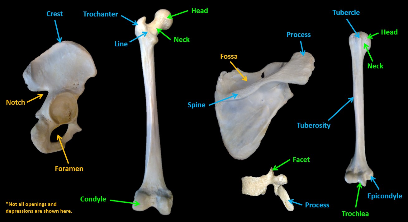
The surface features of bones vary considerably, depending on the function and location in the body. There are three general classes of bone markings:
- Articular surfaces and related features: An articulation is where two bone surfaces come together, forming a joint. These surfaces tend to conform to one another, such as one being rounded and the other cupped, to facilitate the function of the articulation.
- Condyle: Large rounded articular surface
- Facet: Flat articular surface
- Head: Bony expansion carried on a narrow neck
- Neck: Narrowed region next to the head
- Trochlea: smooth spool-shaped surface
- Tendon and/or ligament attachments: These surface features typically project at least a small amount above the surface of the bone or form a rough area on the surface of the bone. In general, their size and shape are an indication of the forces exerted through the attachment to the bone. The larger the attachment site, the greater the force transmitted through the attachment.
- Crest: Narrow prominent ridge of bone
- Epicondyle: A raised area on or above a condyle
- Line: Narrow ridge of bone
- Process: Bony prominence
- Spine: Sharp, slender projection
- Trochanter: Very large, irregularly shaped process
- Tubercle: Small rounded projection or process
- Tuberosity: Large rounded projection, may be roughened
- Opening and depressions: A hole is an opening or groove in the bone that allows blood vessels and nerves to enter the bone. As with the other markings, their size and shape reflect the size of the vessels and nerves that penetrate the bone at these points.
- Fossa: Shallow basin–like depression
- Groove: … a groove
- Sulcus: a larger groove
- Notch: Indentation at the edge of a bone
- Foramen: Round or oval hole through a bone
- Canal: Elongated passage in bone
- Fissure: Slit through bone
- Fovea: a small pit
Muscle Attachments and actions
Muscles attach to bones via tendons. You have already read about the bony prominences that serve as muscle attachment sites. Typically, the proximal attachment site is referred to as the origin, and the distal attachment site is referred to as the insertion. With simple movements in anatomical position, the insertion point, or the bone on which the muscle inserts, moves as the muscle contracts, while the origin remains stable. In some cases, there are multiple origins that converge onto one insertion point, and in other cases, such as the muscles that move the fingers, there may be one origin with multiple insertion points.
Muscles can be grouped based on their primary actions:
- Agonist: the muscle that is acting to complete a particular movement. Several muscles can play a key role in causing a specific movement around a joint. The prime mover is the agonist muscle that is most responsible for completing that specific movement. For example, while several muscles act to flex the hip, the psoas major is the prime mover of hip flexion.
- Antagonist: a muscle with an action that opposes the action of the agonist. For example, the triceps brachii is the antagonist to the muscles that flex the elbow because the triceps brachii acts to extend the elbow.
- Synergist: a muscle that assists with a particular movement. Synergists can function in several different ways. Two agonists can be considered synergists to each other, both contributing to the action occurring at the joint. A synergist can cause secondary motion or a motion at an adjacent joint that allows for a specific motion to occur at the target joint. It can also stabilize a joint and prevent an accessory motion that would inhibit the action of the agonist.
The range of motion and force produced by a muscle is determined by the shape of the muscle.
- The length of the muscle is directly related to the range of motion of that muscle. The longer the muscle fibers are, the greater the range of motion or potential distance the muscle can contract.
- The force a muscle can produce is directly related to the cross-sectional area of the muscle. Muscles with a larger cross-sectional area can produce more force than muscles with a smaller cross-sectional area.
Muscles located on opposite sides of a joint tend to have opposite actions. Muscles are often organized into compartments, and muscles within a compartment typically contribute to the same action. Those in compartments on opposite sides of a joint will perform opposite actions. For example, muscles in the anterior compartment of the forearm will flex the wrist, while muscles in the posterior compartment of the forearm will extend the wrist.
Naming of Skeletal Muscles
Skeletal muscle names tend to tell you something about the muscle itself. You can use the muscle names to your advantage as you are studying the muscles to help you remember the muscle action, location, orientation, or other characteristics of the muscle.
- Some skeletal muscle names refer to the orientation of the muscle fibers. For example, the term “rectus” means straight, so muscles with that term in the name will have straight, long fibers.
- Some muscles have multiple origins, and they are typically referred to as (# term)-ceps. For example, the biceps brachii has two origins or heads, and the triceps has three origins or heads.
- The muscle name can indicate the shape of the muscle. For example, orbicularis means the muscle will form a circle (orbit). The orbicularis oculi muscle circles the eye and functions to close the eye.
- The size of the muscle may be indicated in its name. For example, the gluteus maximus is a large muscle, while the gluteus minimus is smaller. The terms longus and brevis can tell you if the muscle is long or short.
- Muscle names may indicate the location of a muscle. For example, the biceps brachii is located in the arm, while the biceps femoris is located in the thigh. The terms superficialis and profundus will tell you if the muscle is superficial or deep.
- Muscle names may include primary actions of the muscle, such as extensor, flexor, adductor, abductor, or opponens.
- Muscle names can describe the orientation of fibers in the muscle. For example, the external and internal oblique muscles both have fibers that run at an angle.
Some descriptors are only used as necessary. For example, there is no need to include the term “brevis” in the muscle name if there is no “longus” it would be compared to. The flexor pollicis longus muscle flexes the thumb and originates in the forearm. The flexor pollicis brevis muscle also flexes the thumb and is located completely within the hand.
