
Module 24: Muscles and Triangles of the Neck
Learning Objectives:
By the end of this module, students will be able to:
- Explain the organization and distribution of the muscles of the neck.
- Identify the muscles of the anterior neck and explain their function in swallowing/vocalization.
- Identify the major arteries and veins of the neck and explain what tissues or regions are supplied or drained by the main branches.
- Define the triangles of the neck and list the contents of each triangle
Terms to Know
|
The Neck
|
Vasculature of the Head and Neck
|
Muscles That Move the Head
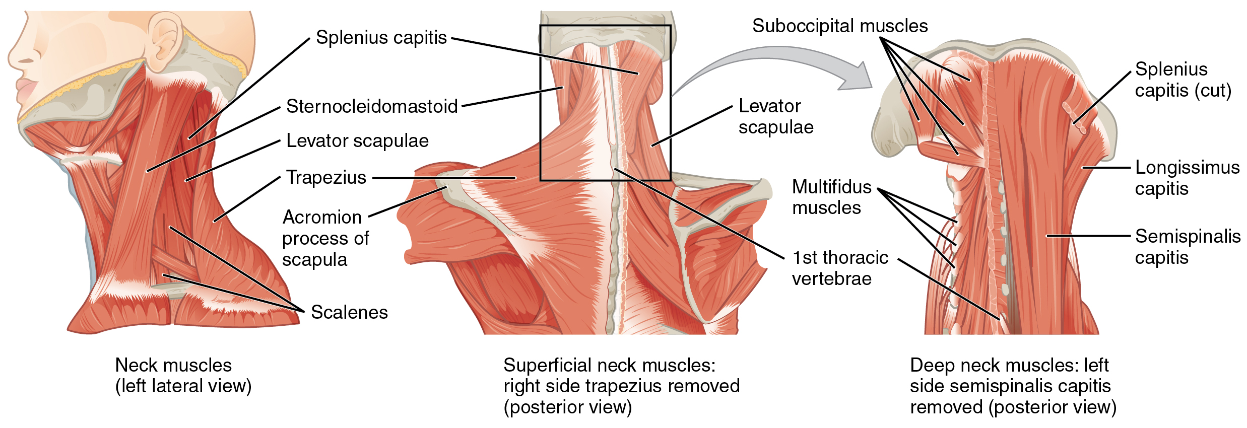
The head, attached to the top of the vertebral column, is balanced, moved, and rotated by the neck muscles. When these muscles act unilaterally, the head rotates. When they contract bilaterally, the neck flexes or extends. The major muscle that laterally flexes and rotates the head is the sternocleidomastoid. In addition, both muscles working together are the flexors of the head. Place your fingers on both sides of the neck and turn your head to the left and to the right. You will feel the movement originate there. This muscle divides the neck into anterior and posterior triangles when viewed from the side.
| Muscles That Move the Head | ||||
|---|---|---|---|---|
| Movement | Target motion direction | Prime mover | Origin | Insertion |
| Neck flexion, rotation, and lateral flexion | Individually: contralateral rotation; bilaterally: flexion | Sternocleidomastoid | Sternum; clavicle | Temporal bone (mastoid process); occipital bone |
| Neck flexion, lateral flexion, rib 1 & 2 elevation | Individually: lateral flexion; bilaterally: flexion, rib elevation | Anterior, Middle, Posterior Scalenes | Transverse process of cervical vertebra | 1st and 2nd ribs |
| Neck extension and rotation | Individually: lateral flexion and ipsilateral rotation; bilaterally: extension | Semispinalis capitis | Transverse and articular processes of cervical and thoracic vertebra | Occipital bone |
| Neck extension, rotation, and lateral flexion | Individually: lateral flexion and ipsilateral rotation; bilaterally: extension | Splenius capitis | Spinous processes of cervical and thoracic vertebra | Temporal bone (mastoid process); occipital bone |
| Neck extension, rotation, and lateral flexion | Individually: lateral flexion and ipsilateral rotation; bilaterally: extension | Longissimus capitis | Transverse and articular processes of cervical and thoracic vertebra | Temporal bone (mastoid process) |
Muscles of the Anterior Neck (Suprahyoid muscles and Infrahyoid muscles)
The muscles of the anterior neck assist in deglutition (swallowing) and speech by controlling the positions of the larynx (voice box), and the hyoid bone, a horseshoe-shaped bone that functions as a solid foundation on which the tongue can move. The muscles of the neck are categorized according to their position relative to the hyoid bone. Suprahyoid muscles are superior to it, and the infrahyoid muscles are located inferiorly.
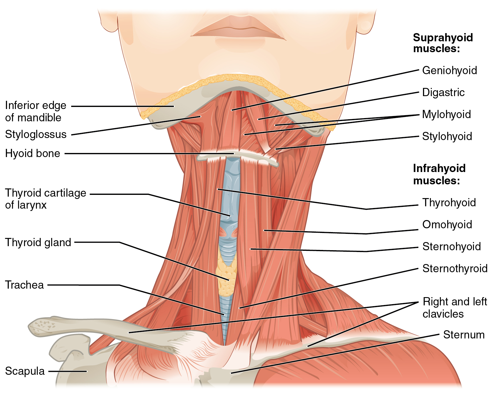
The suprahyoid muscles elevate the hyoid bone, the floor of the mouth, and the larynx during deglutition. These include the digastric muscle, which has anterior and posterior bellies that work to elevate the hyoid bone and larynx when one swallows. The stylohyoid muscle moves the hyoid bone posteriorly, elevating the larynx, and the mylohyoid muscle lifts it and helps press the tongue to the top of the mouth. The geniohyoid depresses the mandible in addition to raising and pulling the hyoid bone anteriorly.
The strap-like infrahyoid muscles generally depress the hyoid bone and control the position of the larynx. The omohyoid muscle, which has superior and inferior bellies, depresses the hyoid bone in conjunction with the sternohyoid and thyrohyoid muscles. The thyrohyoid muscle also elevates the larynx’s thyroid cartilage, whereas the sternothyroid depresses it to create different tones of voice.
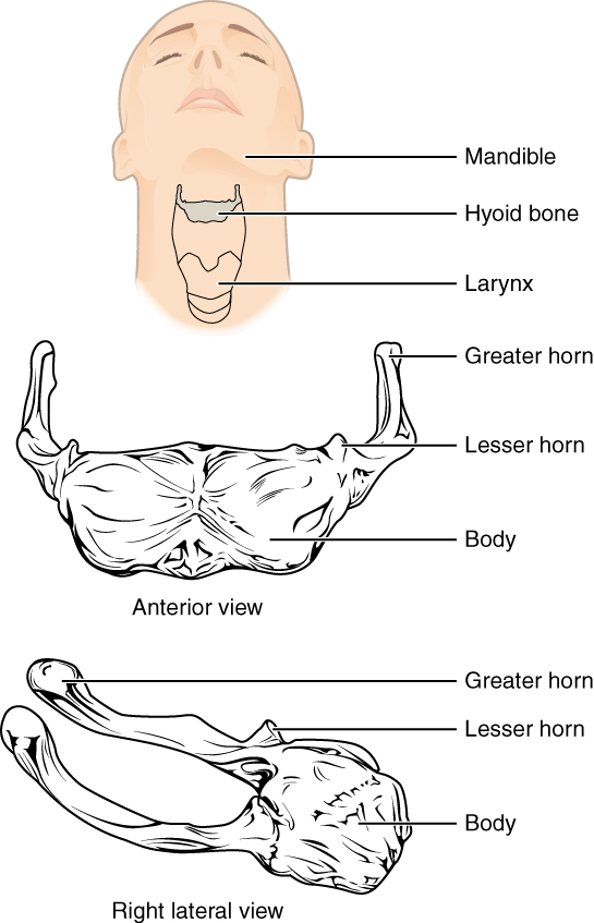
Hyoid Bone
The hyoid bone is an independent bone that does not contact any other bone and thus is not part of the skull. It is a small U-shaped bone located in the upper neck near the level of the inferior mandible, with the tips of the “U” pointing posteriorly. The hyoid serves as the base for the tongue above and is attached to the larynx below and the pharynx posteriorly. The hyoid is held in position by a series of small muscles that attach to it either from above or below. These muscles act to move the hyoid up/down or forward/back. Movements of the hyoid are coordinated with movements of the tongue, larynx, and pharynx during swallowing and speaking.
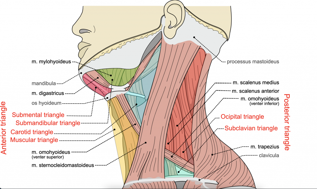
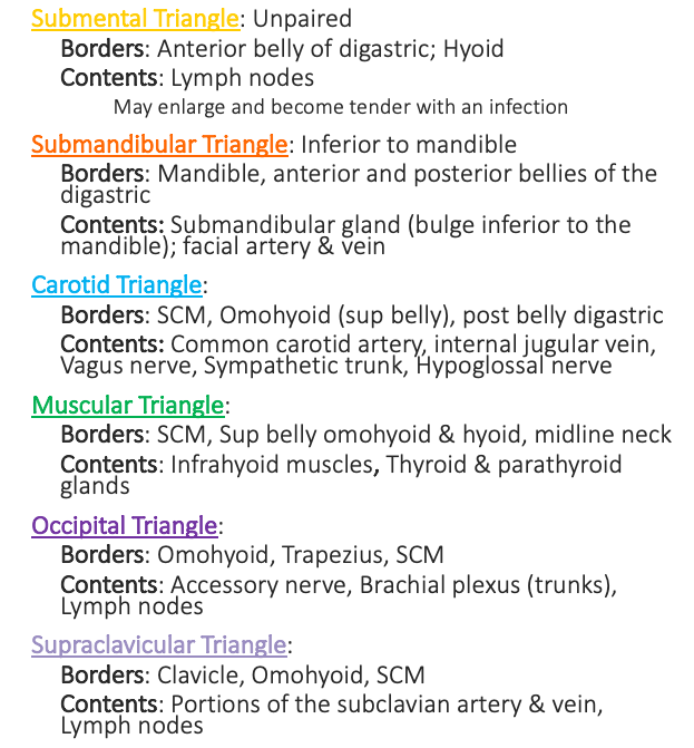
Triangles of the Neck
The triangles of the neck provide an anatomical reference for the location of the contents of the neck. The anterior triangle sits anterior to the sternocleidomastoid muscle and inferior to the mandible. The anterior triangle is subdivided into four smaller triangles, the submental triangle, submandibular triangle, carotid triangle, and muscular triangle. The posterior triangle sits posterior to the sternocleidomastoid muscle and on the lateral region of the neck. The posterior triangle is subdivided into two smaller triangles, the occipital triangle, and the supraclavicular triangle.
Arteries and Veins of the Neck
The arteries and veins of the neck are covered in Module 20 under “Blood Supply to the Brain.” A discussion of the arteries and veins in the head and neck also exists in the lecture videos in Canvas. Students should have an understanding of the location of the arteries and veins of the head and neck, what structures are supplied by the arteries, and which structures are drained by the veins.
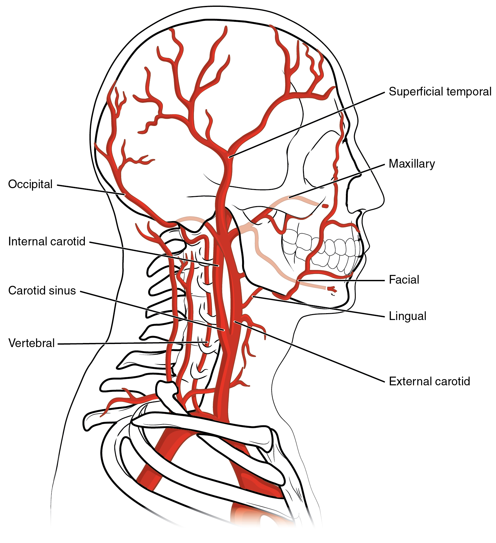
Arteries of the Head and Neck
| Major Arteries of the Head and Neck | |
|---|---|
| Vessel | Description |
| Brachiocephalic artery | Single vessel located on the right side of the body; the first vessel branching from the aortic arch; gives rise to the right subclavian artery and the right common carotid artery; supplies blood to the head, neck, upper limb, and wall of the thoracic region |
| Subclavian artery | The right subclavian artery arises from the brachiocephalic artery while the left subclavian artery arises from the aortic arch; gives rise to the internal thoracic, vertebral, and thyrocervical arteries; supplies blood to the arms, chest, shoulders, back, and central nervous system |
| Vertebral artery | Arises from the subclavian artery and passes through the vertebral foramen through the foramen magnum to the brain; joins with the internal carotid artery to form the arterial circle; supplies blood to the brain and spinal cord |
| Common carotid artery | The right common carotid artery arises from the brachiocephalic artery and the left common carotid artery arises from the aortic arch; each gives rise to the external and internal carotid arteries; supplies the respective sides of the head and neck |
| Internal carotid artery | Arises from the common carotid artery and begins with the carotid sinus; goes through the carotid canal of the temporal bone to the base of the brain; combines with the branches of the vertebral artery, forming the arterial circle; supplies blood to the brain |
| External carotid artery | Arises from the common carotid artery; supplies blood to numerous structures within the face, lower jaw, neck, esophagus, and larynx |
| Superior thyroid artery | Arises from the external carotid artery; supplies blood to the thyroid |
| Lingual artery | Arises from the external carotid artery; supplies blood to the tongue |
| Facial artery | Arises from the external carotid artery; supplies the anterior face |
| Posterior auricular artery | Arises from the external carotid artery; supplies the scalp behind the ear |
| Occipital artery | Arises from the external carotid artery; supplies the occiput, posterior scalp |
| Maxillary artery | Arises from the external carotid artery; supplies the cheek, muscles of mastication, the teeth, and nasal cavity |
| Superficial temporal artery | Arises from the external carotid artery; supplies the lateral and superior scalp |
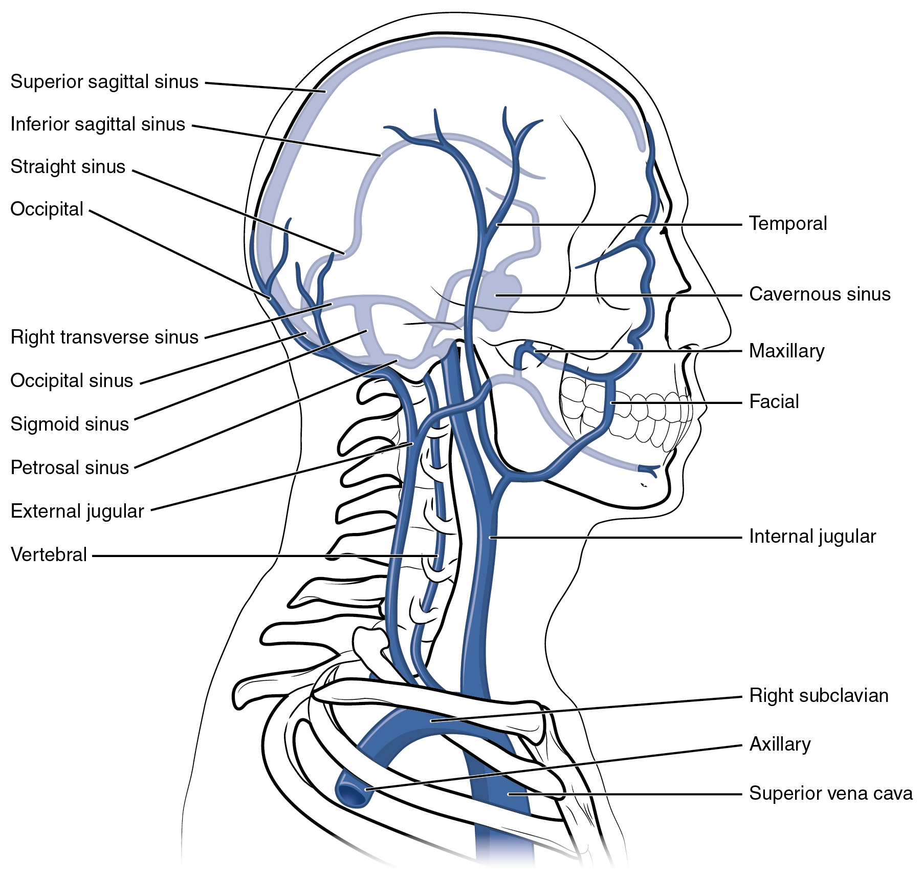
Veins of the Head and Neck
Blood from the brain and the superficial facial veins flow into each internal jugular vein. Blood from the more superficial portions of the head, scalp, and cranial regions, including the temporal vein and maxillary vein, flows into each external jugular vein. Although the external and internal jugular veins are separate vessels, there are anastomoses between them close to the thoracic region. Blood from the internal and external jugular veins empties into the subclavian vein.
| Major Veins of the Head and Neck | |
| Vessel | Description |
| Internal Jugular Vein | Parallel to the common carotid artery, which is more or less its counterpart, and passes through the jugular foramen and canal; primarily drains blood from the brain, receives the superficial facial vein, and empties into the subclavian vein |
| External Jugular Vein | Drains blood from the more superficial portions of the head, scalp, and cranial regions, and leads to the subclavian vein |
| Temporal Vein | Drains blood from the temporal region and flows into the external jugular vein |
| Facial Vein | Drains blood from the anterior face, scalp, and nasal region and flows into the internal jugular vein |
| Maxillary Vein | Drains blood from the maxillary region and flows into the external jugular vein |
