Module 32: Upper Extremity IV – Wrist and Hand
Learning Objectives:
By the end of this class, students will be able to:
- List the bones of the arm and elbow joint.
- Identify the bony and ligamentous structures of the elbow joint.
- Establish relationships between bony landmarks and attachments of muscles/tendons or neurovascular structures in the upper extremities.
- Describe the wrist joint, MP, and IP joints, and infer how the muscle actions will create movement about the joints.
- Review the innervation of muscles of the arm
Terms to Know
|
|
Joints
Muscles of the Hand
|
*NOTE – The table below contains the same information from M30. However, now the only bones presented are the radius, ulna, carpals, metacarpals,
Bones of the Wrist and hand
*NOTE – The table below contains the same information from M30. However, now the only bones presented are the carpals, metacarpals, and phalanges.
| BONES AND BONY MARKINGS OF THE FOREARM WRIST AND HAND |
||
| Carpals | Scaphoid |
Proximal row – most lateral (most common carpal bone fractured) |
| Lunate | Proximal row | |
| Triquetrum | Proximal row | |
| Pisiform | Proximal row – most medial | |
| Trapezium | Distal row – most lateral | |
| Trapezoid | Distal row | |
| Capitate | Distal row | |
| Hamate | Distal row – most medial | |
| Metacarpals | I-V | I = metacarpal of the first ray (thumb), V = metacarpal of fifth ray (pinky) |
| Phalanges | Proximal | Digits I-V have one = 5 total |
| Middle | Digits II-V have one = 4 total | |
| Distal | Digits I-V have one = 5 total | |
Joints of the wrist, hand, and fingers
This content will be covered in the assignment and briefly reviewed in lecture.
| Joints of the Wrist, Hand, and Fingers | |||
| Joint | Bones Articulating | Classification | Motions Available |
| Radiocarpal (Wrist) Joint | Radius and proximal row of carpals | Condyloid (Ellipsoid) Joint | Flexion/Extension Abduction/Adduction Circumduction |
| Carpometacarpal Joint (1st ray only) | Metacarpal I and trapezium | Saddle Joint | Flexion/Extension Abduction/Adduction Circumduction |
| Metacarpophalangeal Joint | Metacarpals and proximal phalanges | Condyloid (Ellipsoid) Joint | Flexion/Extension Abdcution/Adduction Circumduction |
| Interphalangeal Joint | Digit I – proximal and distal phalanges (IP) Digits II-V – proximal and middle phalanges (PIP) Digits II-V – middle and distal phalanges (DIP) |
Hinge Joint | Flexion/Extension |
Wrist, Hand, and Finger Joints
The radiocarpal (wrist) joint is a condyloid joint comprised of the distal radius and the first row of carpal bones. Similar motions to a ball-and-socket joint are available in the wrist. The first metacarpal and trapezium articulate to form the first carpometacarpal joint. The first carpometacarpal joint is a saddle joint. The carpometacarpal joints of digits II-V are more similar to plane joints where only gliding motion occurs. The metacarpophalangeal joints between the proximal phalanges and the distal end of the metacarpals are condyloid joints with similar motions to those seen in the wrists. The interphalangeal joints are hinge joints allowing only flexion and extension to occur between the phalanges. Note the thumb only has an interphalangeal joint, whereas there are proximal and distal interphalangeal joints in digits II-V.
Muscles That Move the Wrist, Hand, and Fingers
Wrist, hand, and finger movements are facilitated by two groups of muscles. The forearm is the origin of the extrinsic muscles of the hand. The palm is the origin of the intrinsic muscles of the hand.
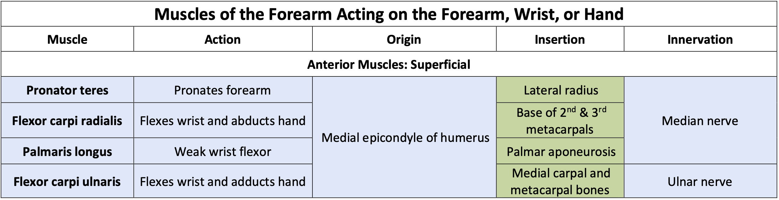
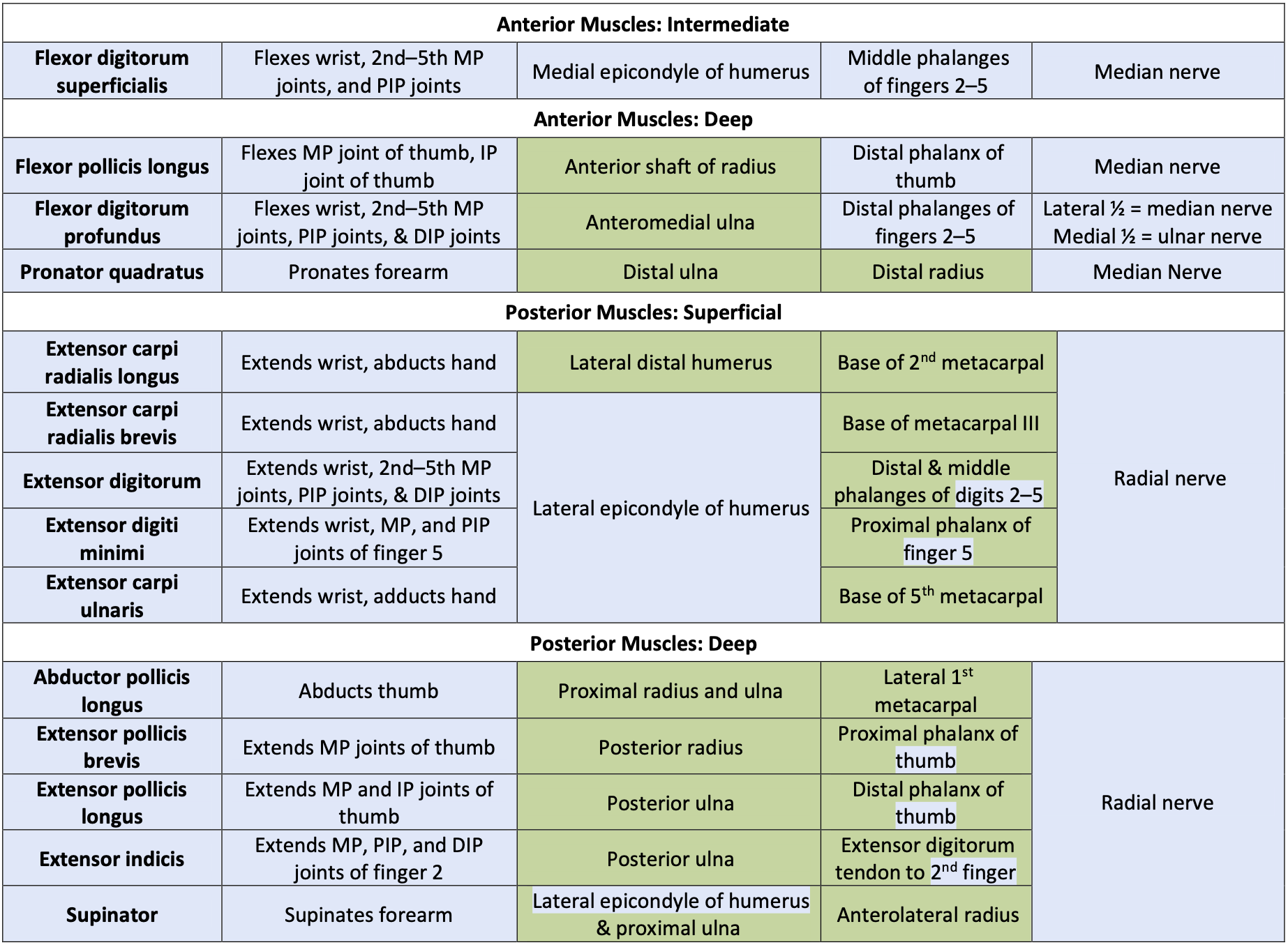
Muscles of the Forearm That Move the Wrists, Hands, and Fingers
This content will be covered in the assignment and reviewed in lecture.
See Module 31 for a discussion of the muscles of the forearm.
Use the image/table below to understand the actions, origins, insertions, and innervation of the muscles that move the elbow and forearm.
*Note: Any box shaded in blue on the muscle chart requires you to know all the information (e.g., action, origin, insertion, and innervation) provided. Areas shaded in green require muscle identification and a general sense of the location of the origin or insertion. (e.g., knowing that the rhomboids originate on the vertebrae, as opposed to the specific numbered vertebrae).
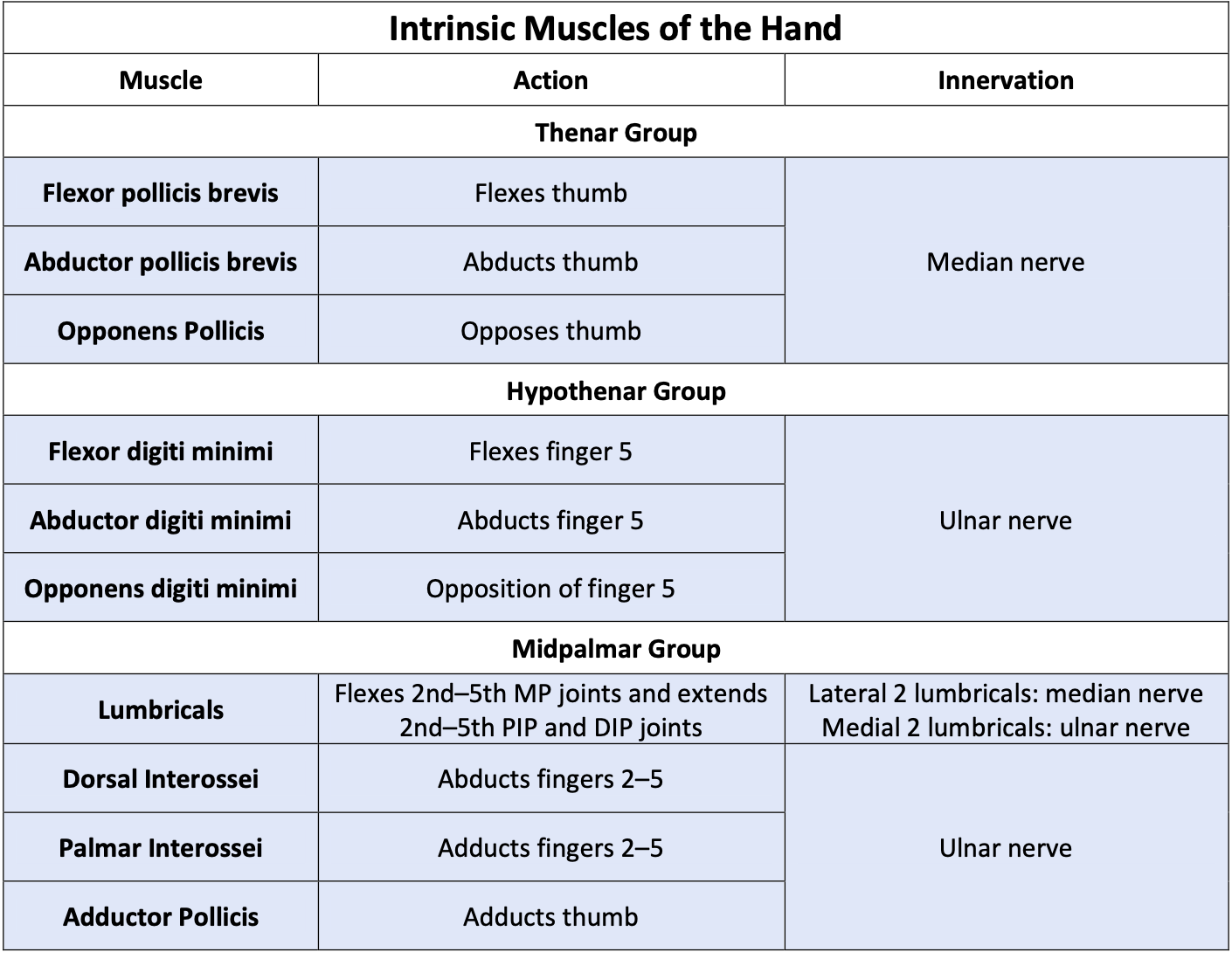
Intrinsic Muscles of the Hand
The intrinsic muscles of the hand both originate and insert within it. These muscles allow your fingers to make precise movements for actions, such as typing or writing. These muscles are divided into three groups. The thenar muscles are on the radial aspect of the palm. The hypothenar muscles are on the medial aspect of the palm. And the intermediate muscles are midpalmar.
The thenar muscles include the abductor pollicis brevis, opponens pollicis, and flexor pollicis brevis. These muscles form the thenar eminence, the rounded contour of the base of the thumb, and all act on the thumb. The movements of the thumb play an integral role in the most precise movements of the hand. The thenar muscles are innervated by the median nerve. The hypothenar muscles include the abductor digiti minimi, flexor digiti minimi brevis, and the opponens digiti minimi. These muscles form the hypothenar eminence, the rounded contour of the little finger, and as such, they all act on the little finger. Finally, the midpalmar muscles act on all the fingers and include the adductor pollicis, the lumbricals, the palmar interossei, and the dorsal interossei. The hypothenar and midpalmar muscles are innervated by the ulnar nerve (**EXCEPT the lateral two lumbricals – they are innervated by the median nerve).
Intrinsic Muscles of the Hand

Use the image/table below to understand the actions, origins, insertions, and innervation of the intrinsic muscles of the hand.
*Note: Any box shaded in blue on the muscle chart requires you to know all the information (e.g., action, origin, insertion, and innervation) provided. You do not need to know the origins or insertions for the intrinsic muscle of the hand.