
Module 27: Cranial Nerve Review
Learning Objectives:
By the end of this class, students will be able to:
- List the names and locations of the 12 cranial nerves.
- Describe the general pathway and principal functions of each cranial nerve.
- List which cranial nerves carry autonomic fibers and identify the ganglia associated with those cranial nerves.
- Describe the effects of autonomic innervation on various effectors in the head and neck.
Terms to Know
|
Cranial Nerves
*Covered only in lecture, not in this text |
Neural Modalities
Autonomics of Head and Neck
|
The assignment for this module will review all information from the posted cranial nerve slides and this eReader module. The cranial nerves will be covered throughout this unit, but students should begin studying this information early, as it serves as foundational information for the unit. This module and class will review information that you have learned about the cranial nerves, and highlight a few important points that were not mentioned elsewhere in this unit.
Neural Modalities
This information is reviewed in the Brain Part I lecture.
A neural modality is the type of information carried in a nerve. We will discuss five neural modalities that are carried in cranial nerves. Some carry just one, while others carry all five modalities. You should know which modalities originate and are carried in each cranial nerve. (Note: we only discuss resident modalities, meaning modalities that originate in a given cranial nerve. Some cranial nerves pick up fibers with other modalities from other cranial nerves along their path, but you are not responsible for knowing that information.)
- General somatic sensory: Sense of touch, proprioception, pain, and temperature
- General somatic motor: Conscious motor innervation of skeletal muscles. ***You may see special motor on some slides. Special motor fibers also innervate skeletal muscles, but they develop in a different way. We will group special motor with somatic motor in this course.
- General visceral sensory: Unconscious sensation from our visceral structures (e.g. organs, glands, blood vessels, monitoring blood oxygen content, etc.)
- General visceral motor: Unconscious motor innervation to cardiac or smooth muscle and glands
- Special sensory: vision, audition (hearing), taste, vestibulation, and smell
Cranial Nerves Review
This section will provide a brief review of the cranial nerves. This is covered in the lecture slides, and some of it is also reviewed in lecture.
The cranial nerves are primarily responsible for the sensory and motor functions of the head and neck (except for thoracic and abdominal organs innervated by the vagus nerve). There are twelve cranial nerves, which are designated CN I through CN XII for “Cranial Nerve,” using Roman numerals. CN I is associated with the telencephalon, and CN II is associated with the diencephalon. The other ten cranial nerves originate from the brainstem.
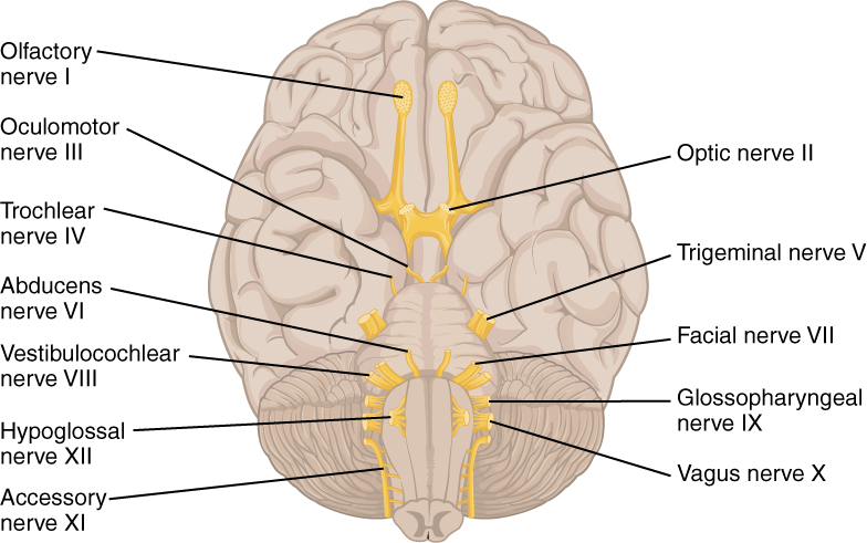
CN I: Olfactory Nerve
- Modality
- Special sensory: Olfaction (smell)
- Skull passageway: Cribriform plate foramina of the ethmoid bone
- Other Notes: Synapses in the olfactory bulbs. Cranial nerve one is only the short nerve traveling from the olfactory mucosa through the cribriform plate foramina to the olfactory bulb. You cannot clearly see the olfactory nerves on a brain that has been removed from the skull, as they have been cut. (The image above is trying to point to the olfactory nerves synapsing with the olfactory bulb, represented by small darker dots on the olfactory bulb.)
CN II: Optic Nerve
- Modality
- Special sensory: Vision
- Skull passageway: Optic canal
- Other Notes: This is technically a tract of the brain that is covered in meninges. However, it does not have the name optic tract until after nasal fibers cross in the optic chiasm.
CN III: Oculomotor Nerve
- Modality
- Somatic motor: Innervates four of the six extrinsic eye muscles, superior rectus, inferior rectus, medial rectus, and inferior oblique. Also innervates one muscle of facial expression that is located in the orbit, the levator palpebrae superioris.
- Visceral motor: Sphincter pupillae and the ciliary muscle
- Preganglionic parasympathetic fibers synapse in the ciliary ganglion and then travel to the sphincter pupillae in the iris and ciliary muscle.
- Skull passageway: Superior orbital fissure
CN IV: Trochlear Nerve
- Modality
- Somatic motor: Innervates the superior oblique, an extrinsic eye muscle
- Skull passageway: Superior orbital fissure
- Other Notes: Gets its name because the tendon of the muscle it innervates travels through a trochlea, or pulley, at the superior medial aspect of the orbit
CN V: Trigeminal Nerve
- Modality
- Somatic motor: Innervates the four muscles of mastication as well as the myelohyoid, anterior belly of the digastric, tensor tympani, and tensor veli palatini muscles. These are only innervated by the mandibular division.
- Somatic sensory: Sense of touch, temperature, proprioception, and pain from the face
-
V1: cornea, nose, forehead, anterior scalpV2: nasal mucosa, palate, gums, cheekV3: anterior 2/3 tongue, meninges, skin of the chin, lower jaw, lower teeth, part of the ear
-
- Skull passageway: Superior orbital fissure for the ophthalmic division (you do not need to know the foramen for the maxillary or mandibular divisions in this course)
- Other Notes: Splits into three divisions, the ophthalmic (V1), maxillary (V2), and mandibular (V3). Sensory axons enter the cranium and synapse at the trigeminal ganglion, which is like the dorsal root ganglion for the trigeminal nerve.
CN VI: Abducens Nerve
- Modality
- Somatic motor: Innervates the lateral rectus, an extrinsic eye muscle
- Skull passageway: Superior orbital fissure
CN VII: Facial Nerve
- Modality
- Special sensory: Taste from the anterior 2/3 of the tongue
- Visceral motor: Innervates and promotes secretion from several glands of the head and neck
- Some preganglionic parasympathetic fibers synapse in the pterygopalatine ganglion. Postganglionic fibers leaving this ganglion travel to the lacrimal gland and other glands scattered in the mucosa of the nasal cavity, oral cavity, palate, and pharynx
- Some preganglionic parasympathetic fibers synapse in the submandibular ganglion. Postganglionic fibers leaving this ganglion travel to the submandibular and sublingual salivary glands.
- Visceral sensory: Monitoring the secretion from the glands above
- Somatic sensory: Sense of touch from a small area behind the ear
- Somatic motor: Innervates the muscles of facial expression (except levator palpebrae superioris, which is innervated by CN III) as well as the posterior belly of the digastric, stylohyoid, and stapedius muscles
- Skull passageway: Enters the skull through the internal acoustic meatus. The branch that innervates the muscles of facial expression exits the skull and enters the face via the stylomastoid foramen
CN VIII: Vestibulocochlear Nerve
- Modality
- Special sensory:
- Cochlear branch: Audition (hearing)
- Vestibular branch: Equilibrium and balance
- Special sensory:
- Skull passageway: Internal acoustic meatus
CN IX: Glossopharyngeal Nerve
- Modality
- Special sensory: Taste from the posterior 1/3 of the tongue
- Somatic sensory: Sense of touch, temperature, pain from the pharynx
- Visceral motor: Innervates and promotes secretion from the parotid gland, a salivary gland
- Preganglionic parasympathetic fibers synapse in the otic ganglion, then postganglionic fibers travel to the parotid gland
- Visceral sensory: Monitors secretion from the parotid gland, also monitors oxygen and carbon dioxide levels in the blood via the carotid bodies, which are located at the point where the common carotid artery branches into the internal and external carotid arteries
- Somatic motor: Innervates the stylopharyngeus muscle
- Skull passageway: Jugular foramen
CN X: Vagus Nerve
- Modality
- Special sensory: Taste from the base of the tongue, epiglottis, upper pharynx
- Visceral motor: Innervates the heart, pharynx, larynx, trachea, lungs, midgut and hindgut organs
- Visceral sensory: Monitors contraction of smooth and cardiac muscle and secretion from glands in the organs above
- Somatic sensory: Sense of touch, pain, and temperature from the external acoustic meatus, eardrum, laryngopharynx, larynx
- Somatic motor: Muscles of the pharynx, including the pharyngeal constrictors, muscles of the larynx and palate
-
Recurrent laryngeal nerve: Branches in the thorax, left loops under the arch of the aorta, returns to neck to innervate laryngeal muscles
-
- Skull passageway: Jugular foramen
CN XI: Accessory Nerve
- Modality
- Somatic motor: Innervates the trapezius and sternocleidomastoid muscles
- Skull passageway: Jugular foramen
- Other Notes: Some consider this to be a branch of the vagus nerve. We will consider it a separate nerve in this course. It is also sometimes called the spinal accessory nerve.
CN XII: Hypoglossal Nerve
- Modality
- Somatic motor: Innervates the intrinsic and extrinsic muscles of the tongue
- Skull passageway: Hypoglossal canal
