
Module 29: Upper Extremity I – Neurovasculature
Learning Objectives:
By the end of this class, students will be able to:
- Identify the nerves of the brachial plexus.
- List the terminal branches.
- List the named nerves originating from the brachial plexus.
- Identify the components of the brachial plexus.
- Differentiate a root, trunk, division, cord, and terminal branch
- Explain proximal to distal gradient of innervation.
- Identify the locations of the dermatomes of the upper extremity.
- Identify the spinal nerve root associated with the dermatome.
- Identify the myotomes of the upper extremity.
- List the motion associated with each spinal nerve root.
- Identify the sensory innervation of each terminal branch of the brachial plexus.
- Identify the muscles innervated by each terminal branch of the brachial plexus.
- Sketch and interpret the brachial plexus.
- List the major blood vessels of the upper extremities and describe their location.
- Identify the muscles and/or regions supplied or drained by the major blood vessels of the upper extremities.
Terms to Know
|
Vasculature
|
Brachial Plexus
|
Arteries Serving the Upper Limbs
This content will be briefly covered in the assignment and built upon in lecture.
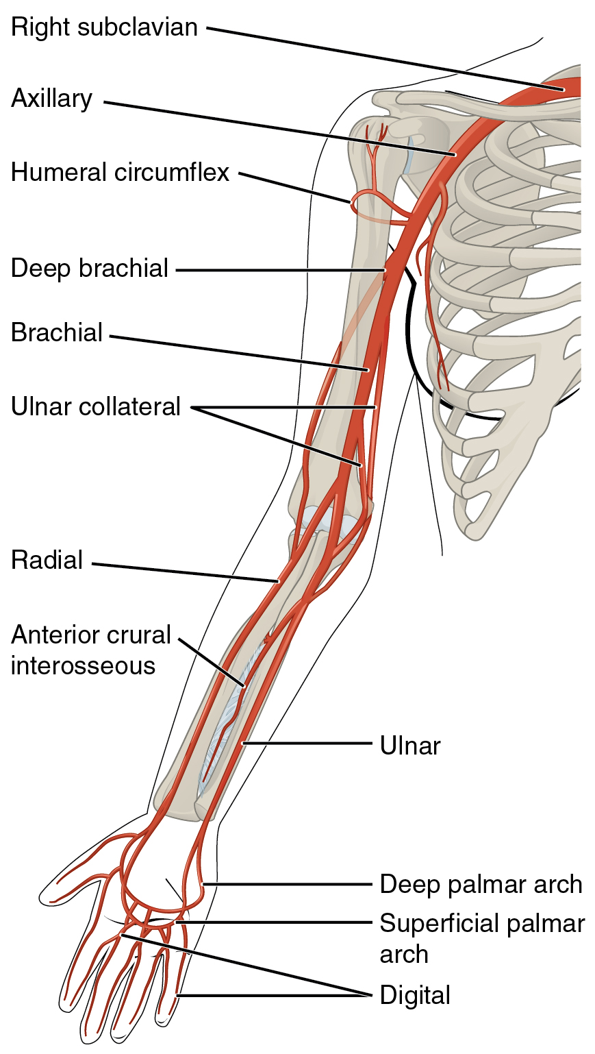
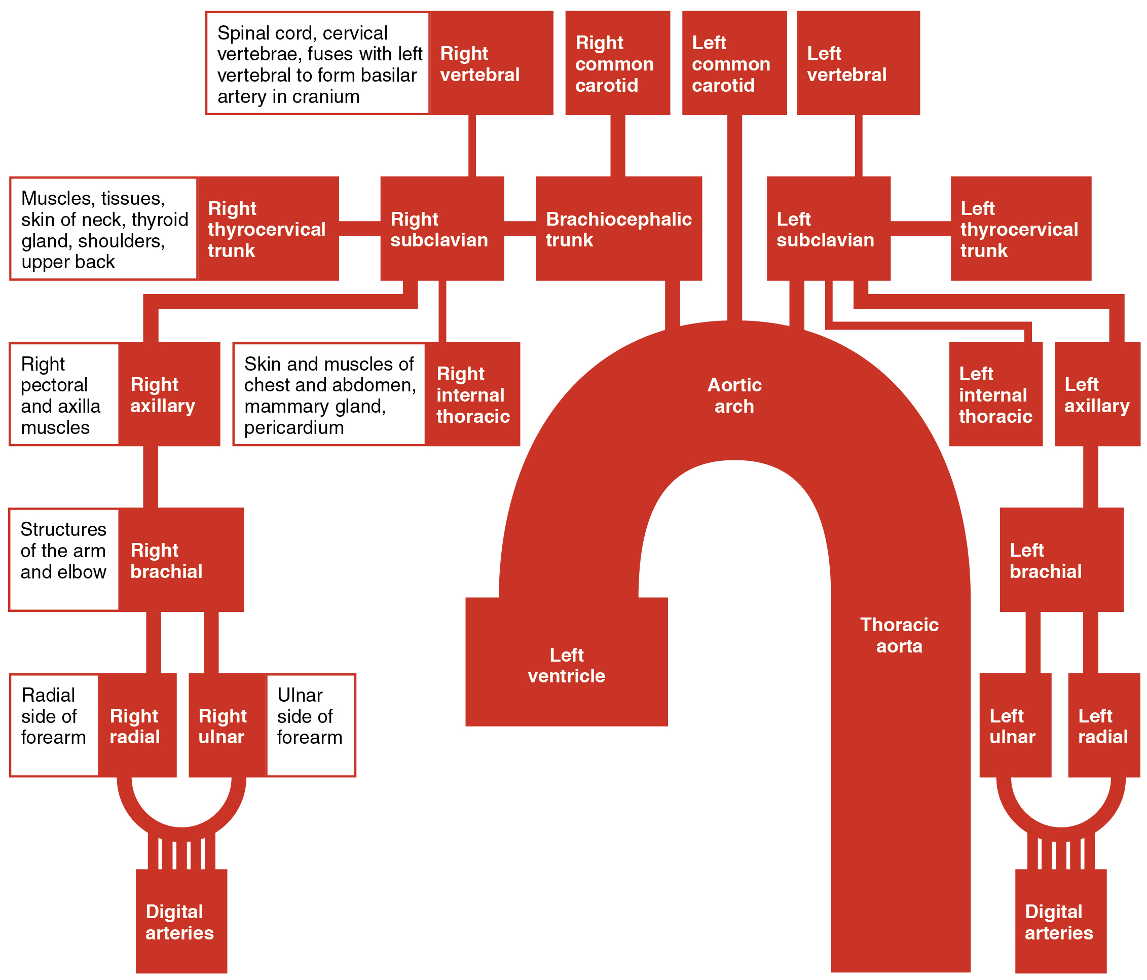
The subclavian artery has a major branch, the thyrocervical trunk, supplying the neck, shoulders, and upper back. The thyrocervical trunk has two major branches, the suprascapular artery supplying the supraspinatus and infraspinatus, and the dorsal scapular artery supplying the levator scapulae, rhomboids, and trapezius muscles. As the subclavian artery exits the thorax into the axillary region, it is renamed the axillary artery. The axillary artery comprises three parts, each with branches supplying different parts of the axilla and pectoral muscles. Although it does branch and supply blood to the axillary region, most of the vessel continues into the upper arm, or brachium, and becomes the brachial artery. The brachial artery supplies blood to much of the brachial region, including a branch called the deep brachial artery, which provides blood to the posterior surface of the arm. As the brachial artery approaches the cubital fossa, it bifurcates into the radial and ulnar arteries, which continue into the forearm, or antebrachium. The radial artery and ulnar artery parallel their namesake bones, giving off smaller branches until they reach the wrist. In the hand, they fuse to form the superficial and deep palmar arches that supply blood to the hand and the digital arteries that supply blood to the digits.
| ARTERIES SERVING THE UPPER LIMBS | |
|---|---|
| Vessel | Description |
| Axillary artery | Continuation of the subclavian artery as it penetrates the body wall and enters the axillary region; comprises three parts; supplies blood to the axillary region and pectoral muscles; most of the vessel continues into the brachium and becomes the brachial artery. |
| Brachial artery | Continuation of the axillary artery in the brachium; supplies blood to much of the brachial region; gives off several smaller branches that provide blood to the posterior surface of the arm in the region of the elbow; bifurcates into the radial and ulnar arteries at the cubital fossa. |
| Radial artery | Formed at the brachial artery’s bifurcation; parallels the radius; gives off smaller branches until it reaches the carpal region where it fuses with the ulnar artery to form the superficial and deep palmar arches; supplies blood to the lower arm and carpal region. |
| Ulnar artery | Formed at the brachial artery’s bifurcation; parallels the ulna; gives off smaller branches until it reaches the carpal region where it fuses with the radial artery to form the superficial and deep palmar arches; supplies blood to the lower arm and carpal region. |
| Palmar arches (superficial and deep) | Formed from anastomosis of the radial and ulnar arteries; supply blood to the hand and digital arteries. |
| Digital arteries | Formed from the superficial and deep palmar arches; supply blood to the digits. |
Veins Draining the Upper Limbs
This content will be briefly covered in the assignment and built upon in lecture.
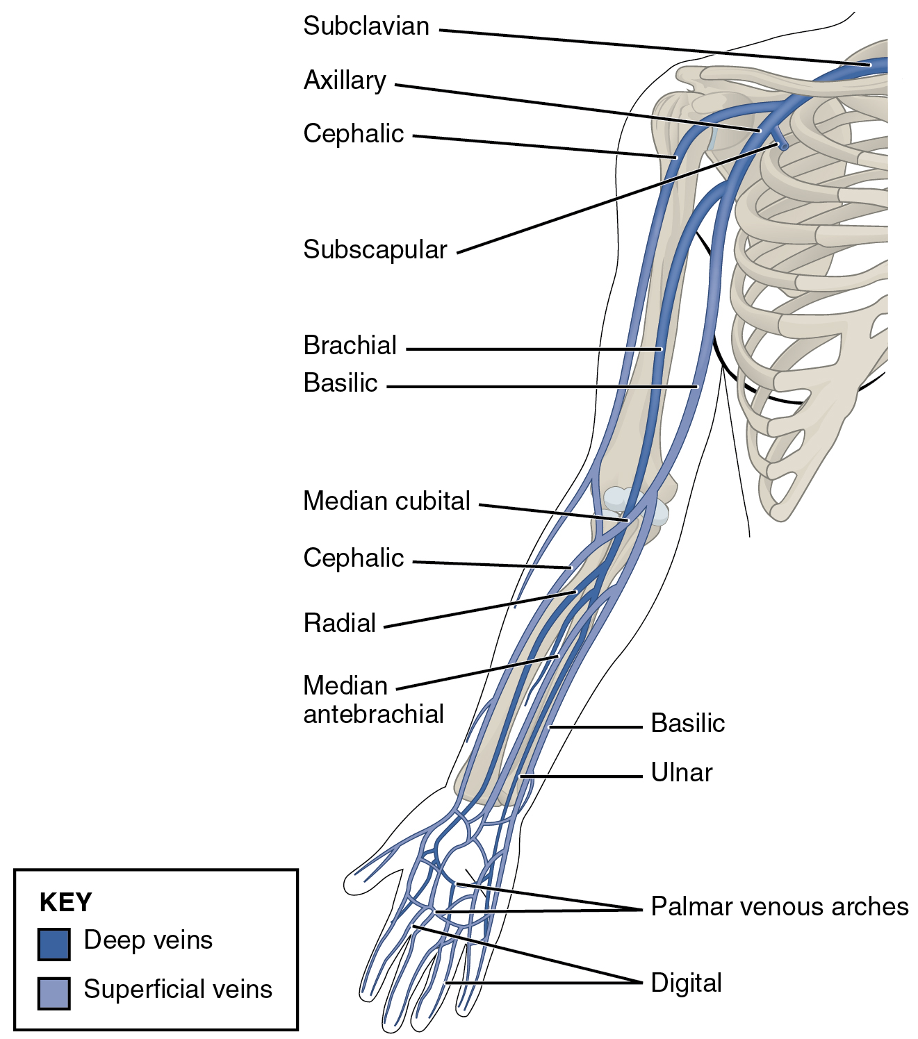
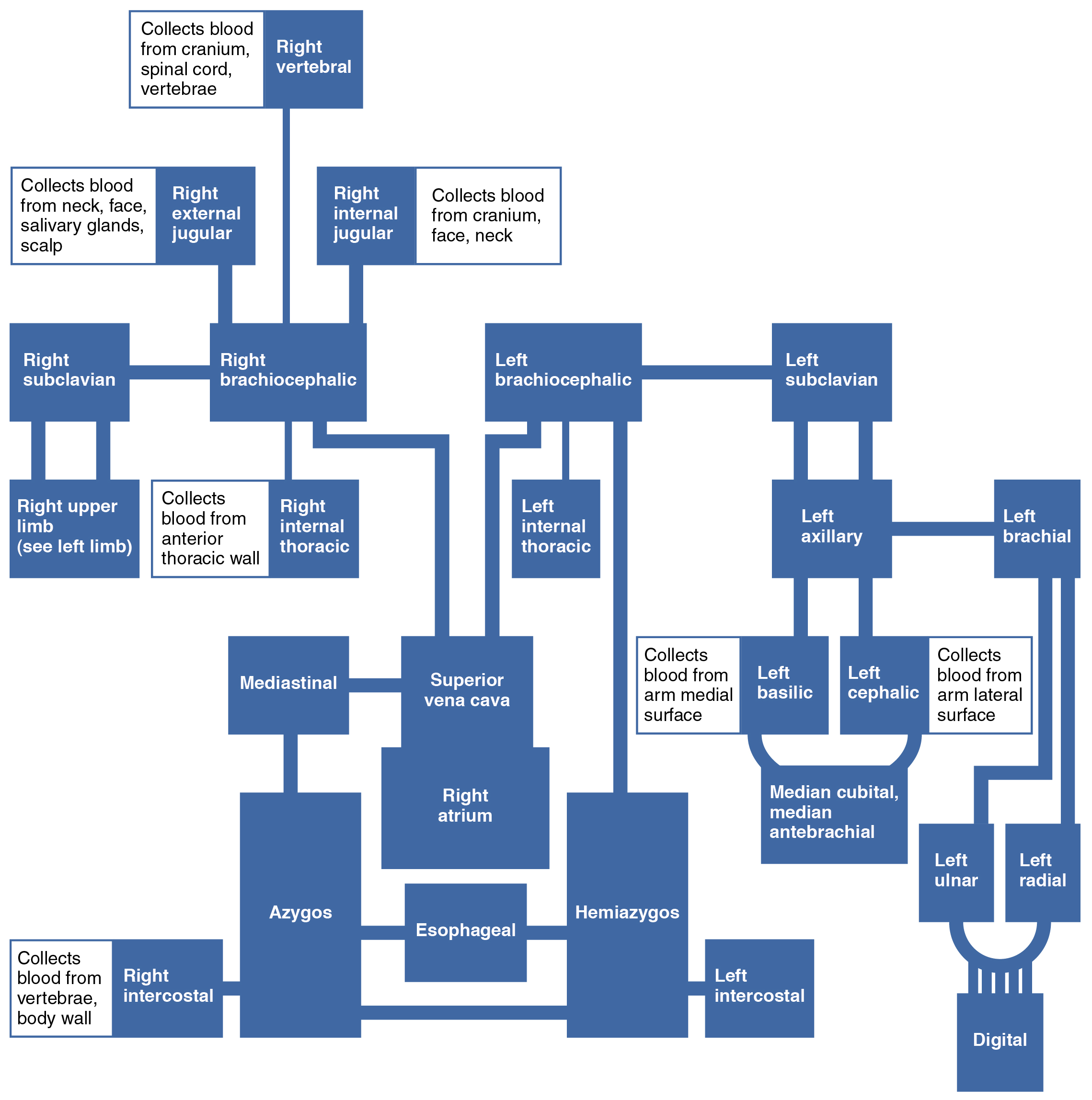
The digital veins in the fingers come together in the hand to form the palmar venous arches. From here, the veins come together to form the radial vein, the ulnar vein, and the median antebrachial vein. The radial vein and the ulnar vein parallel the forearm bones and join together at the antebrachium to form the brachial vein, a deep vein that flows into the axillary vein in the brachium.
The median antebrachial vein parallels the ulnar vein, is more medial in location, and joins the basilic vein in the forearm. As the basilic vein reaches the antecubital region, it gives off a branch called the median cubital vein that crosses at an angle to join the cephalic vein. The median cubital vein is the most common site for drawing venous blood in humans. The basilic vein continues through the arm medially and superficially to the axillary vein.
The cephalic vein begins in the antebrachium and drains blood from the superficial surface of the arm into the axillary vein. It is extremely superficial and easily seen along the surface of the biceps brachii muscle in individuals with good muscle tone and those without excessive subcutaneous adipose tissue in the arms.
The subscapular vein drains blood from the subscapular region and joins the cephalic vein to form the axillary vein. As it passes through the body wall and enters the thorax, the axillary vein becomes the subclavian vein.
| VEINS OF THE UPPER LIMBS | |
|---|---|
| Vessel | Description |
| Digital veins | Drain the digits and lead to the palmar arches of the hand and dorsal venous arch of the foot |
| Palmar venous arches | Drain the hand and digits, and lead to the radial vein, ulnar veins, and the median antebrachial vein |
| Radial vein | Vein that parallels the radius and radial artery; arises from the palmar venous arches, and leads to the brachial vein. |
| Ulnar vein | Vein that parallels the ulna and ulnar artery; arises from the palmar venous arches, and leads to the brachial vein. |
| Brachial vein | Deeper vein of the arm that forms from the radial and ulnar veins in the lower arm; leads to the axillary vein. |
| Basilic vein | Superficial vein of the arm that arises from the median antebrachial vein, intersects with the median cubital vein, parallels the ulnar vein, and continues into the upper arm; along with the brachial vein, it leads to the axillary vein. |
| Median cubital vein | Superficial vessel located in the antecubital region that links the cephalic vein to the basilic vein in the form of a v; a frequent site from which to draw blood |
| Cephalic vein | Superficial vessel in the upper arm; leads to the axillary vein. |
| Axillary vein | The major vein in the axillary region; drains the upper limb and becomes the subclavian vein. |
The Brachial Plexus
The content on nerve roots and terminal branches will be covered in the assignment. The rest will be covered in lecture.
The brachial plexus is a network of nerves innervating the upper extremities. Exiting from the cervical and upper thoracic vertebrae, the brachial plexus travels within the axilla towards the target tissues.
- Roots
- The anterior rami of C5-T1 form the roots of the brachial plexus, which unite to form trunks.
- Trunks
- The superior trunk includes nerve roots C5 and C6.
- The middle trunk is a continuation of nerve root C7.
- The inferior trunk includes nerve roots C8 and T1.
- The trunks subsequently divide into anterior and posterior divisions.
- Divisions
- The anterior divisions will supply the anterior arm, forearm, and hand.
- The posterior divisions will innervate the posterior arm, forearm, and hand.
- Cords
- The anterior divisions from the superior and middle trunk will form the lateral cord.
- The anterior division from the inferior trunk will continue as the medial cord.
- The posterior divisions from each trunk will form the posterior cord.
- The cords are named in their relation to the axillary artery.
- The lateral and medial cords are lateral and medial, respectively, to the axillary artery.
- The posterior cord is posterior to the axillary artery.
- Terminal Branches
- Five terminal branches are emerging from the cords, the axillary nerve, median nerve, musculocutaneous nerve, radial nerve, and ulnar nerve.
Many named nerves arise from the brachial plexus. The table below highlights the named nerves arising from the brachial plexus, the nerve roots comprising the nerves, and where applicable, the muscles innervated by the nerves and the sensory distribution of the nerves.
| NERVES OF THE BRACHIAL PLEXUS | |||
| Named Nerve of the Brachial Plexus | Nerve Root(s) | Muscle(s) Innervated | Sensory Distribution |
| Dorsal Scapular Nerve | C5 | Levator Scapulae Rhomboid Minor Rhomboid Major |
– |
| Long Thoracic Nerve | C5-C7 | Serratus Anterior | – |
| Nerve to Subclavius | C5-C6 | Subclavius | – |
| Suprascapular Nerve | C5-C6 | Supraspinatus Infraspinatus |
– |
| Lateral Pectoral Nerve | C5-C7 | Pectoralis Major | – |
| Upper Subscapular Nerve | C5-C6 | Subscapularis | – |
| Lower Subscapular Nerve | C5-C6 | Subscapularis Teres Major |
– |
| Thoracodorsal Nerve | C6-C8 | Latissimus Dorsi | – |
| Medial Pectoral Nerve | C8-T1 | Pectoralis Major Pectoralis Minor |
– |
| Medial Brachial Cutaneous | C8-T1 | – | Medial skin of the arm |
| Medial Antebrachial Cutaneous | C8-T1 | – | Medial skin of the forearm |
| Axillary Nerve | C5-C6 | Deltoid Teres Minor |
Anterolateral to posterolateral shoulder |
| Radial Nerve | C5-T1 | Posterior Arm & Forearm Triceps Brachii Brachioradialis Supinator Extensor Carpi Ulnaris Extensor Digiti Minimi Extensor Digitorum Extensor Carpi Radialis Longus Extensor Carpi Radialis Brevis Abductor Pollicis Longus Extensor Pollicis Longus Extensor Pollicis Brevis Extensor Indicis |
Posterior arm and forearm, posterolateral hand |
| Musculocutaneous Nerve | C5-C7 | Anterior Arm Coracobrachialis Biceps Brachii Brachialis |
Lateral forearm |
| Median Nerve | C6-T1 | Most of the Anterior Forearm and Lateral Hand Pronator Teres Flexor Carpi Radialis Palmaris Longus Flexor Digitorum Superficialis Flexor Digitorum Profundus (lateral half) Flexor Pollicis Longus Pronator Quadratus Lumbricals (lateral two) Opponens Pollicis Abductor Pollicis Brevis Flexor Pollicis Brevis |
Anterolateral hand, thumb, distal posterior digits 2 & 3 |
| Ulnar Nerve | C8-T1 | Medial Anterior Forearm & Most of the Hand Flexor Carpi Ulnaris Flexor Digitorum Profundus (medial half) Adductor Pollicis Dorsal Interossei Palmar Interossei Lumbricals (medial two) Opponens Digiti Minimi Abductor Digiti Minimi Flexor Digiti Minimi |
Medial hand anterior and posterior |
Proximal to Distal Gradient of Innervation
This content will be covered in lecture.
Nerves supplied by the superior anterior rami (C5-C6) will innervate the most proximal muscles. Nerves with intermediate rami fibers (C6-C8) will innervate the muscles acting on the elbow and wrist. The nerves with the most inferior rami (C8-T1) will innervate the most distal (hand) muscles.
A deeper discussion on the brachial plexus, dermatomes, myotomes, named nerves, and the terminal branches will be found in videos on Canvas.
