
Module 23: Skull and Muscles of the Face
Learning Objectives:
By the end of this module, students will be able to:
- List the cranial and facial bones of the skull and identify key anatomical features/landmarks.
- Name and describe/locate the sutures between cranial bones.
- Identify the various foramen of the skull and list what travels through them.
- Describe the major muscles involved in facial expression and their functions
Terms to Know
|
Skull
|
Muscles of Facial Expression
|
*NOTE – Most of the reading about the cranial and facial bones is very in-depth. Remember – the learning objective is to list the bones and identify key features/landmarks (identified above for each bone).
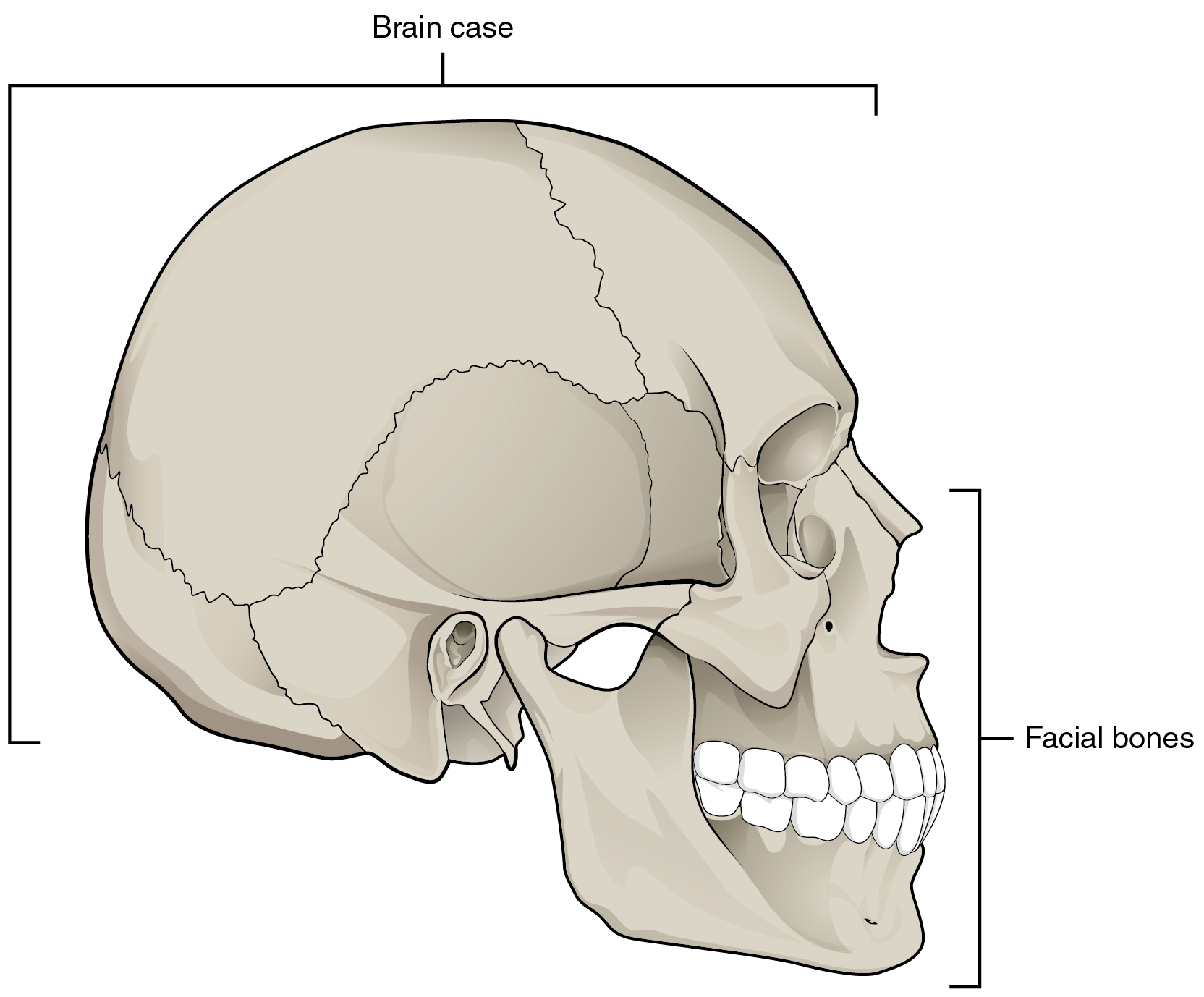
The cranium (skull) is the skeletal structure of the head that supports the face and protects the brain. It is subdivided into the facial bones and cranial bones. The facial bones underlie the facial structures, form the nasal cavity, enclose the eyeballs, and support the teeth of the upper and lower jaws. The rounded cranium of the cranial bones surrounds and protects the brain and houses the middle and inner ear structures.
Anterior View of Skull
The anterior skull consists of the facial bones and provides the bony support for the eyes and structures of the face. This view of the skull is dominated by the openings of the orbits and the nasal cavity. Also seen are the upper and lower jaws, with their respective teeth.
The orbit is the bony socket that houses the eyeball and muscles that move the eyeball or open the upper eyelid. The upper margin of the anterior orbit is the supraorbital margin.
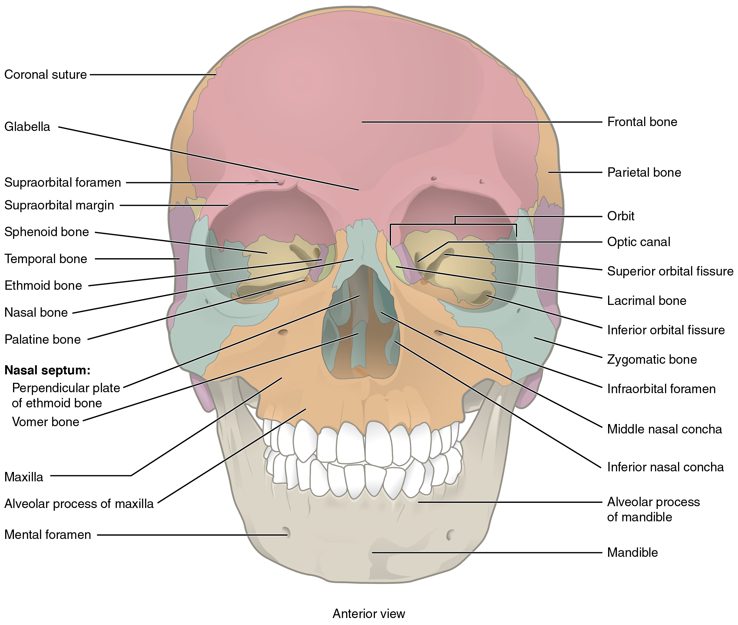
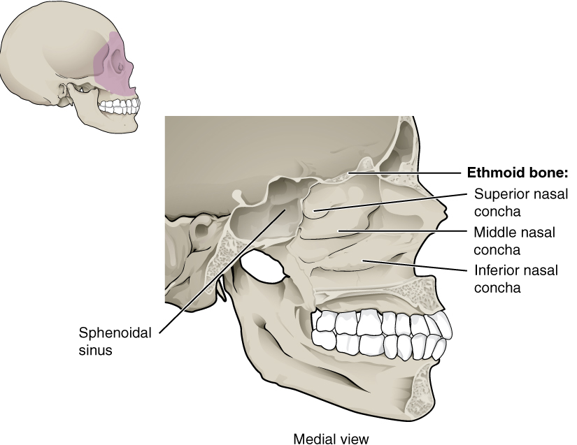
Inside the nasal area of the skull, the nasal cavity is divided into halves by the nasal septum. The upper portion of the nasal septum is formed by the perpendicular plate of the ethmoid bone and the lower portion is the vomer bone. Each side of the nasal cavity is triangular in shape, with a broad inferior space that narrows superiorly. The nasal concha (superior – part of the ethmoid, middle – part of the ethmoid, and inferior) are found in the nasal cavity.
Lateral View of Skull
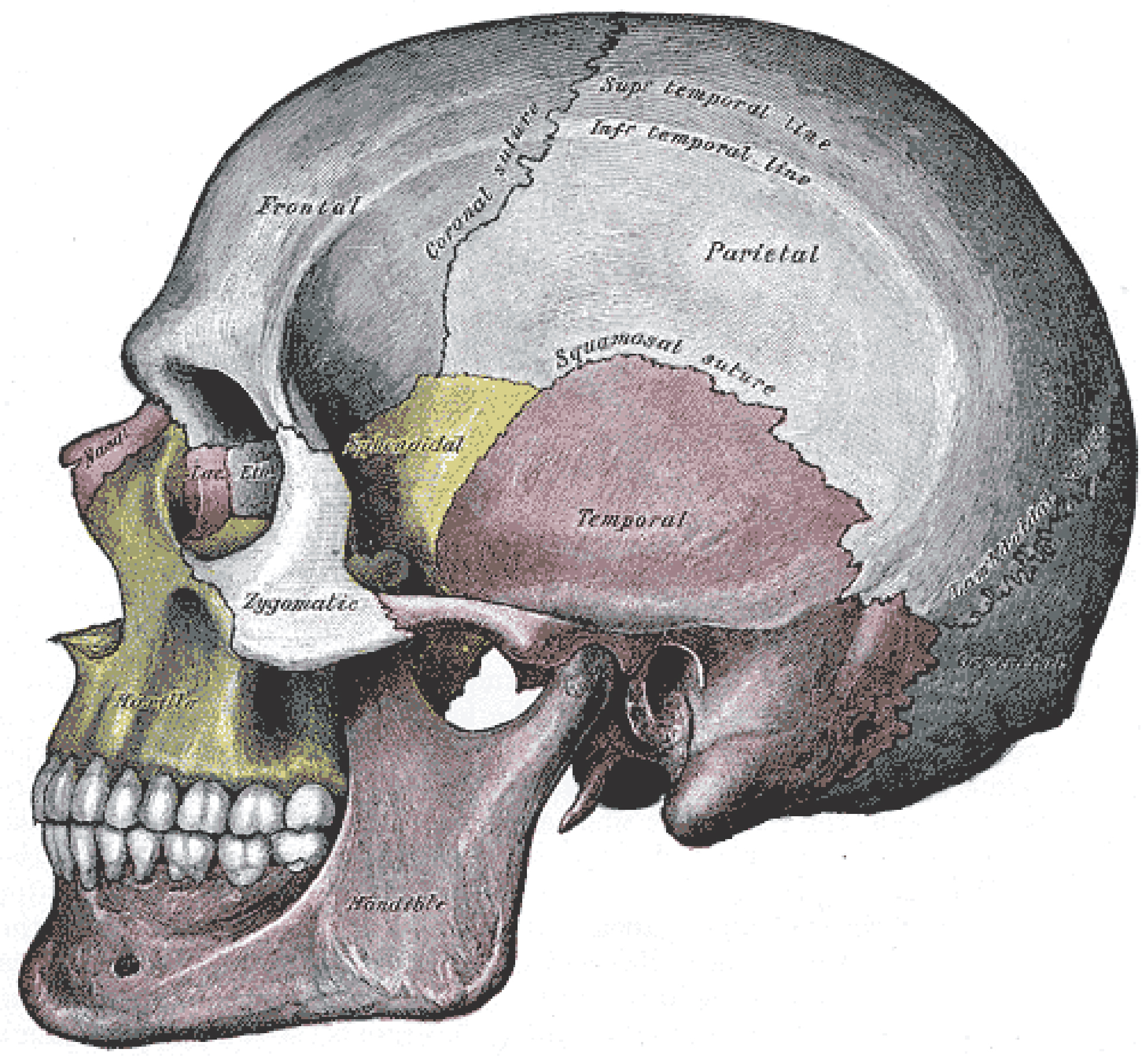
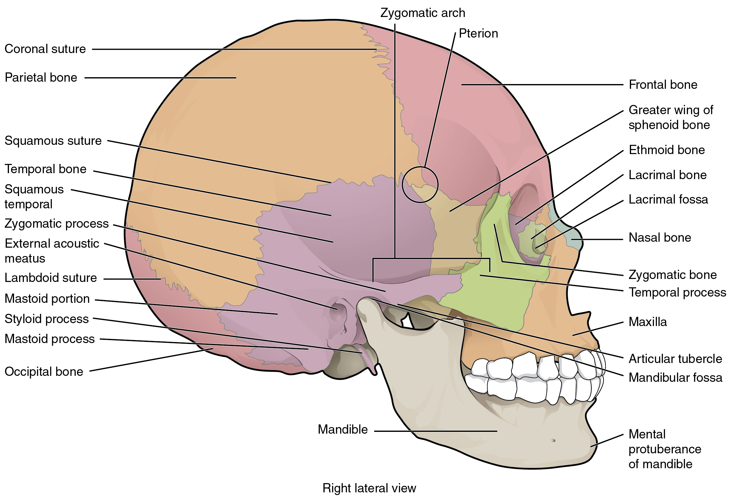
A view of the lateral skull is dominated by the large, rounded cranium above and the upper and lower jaws with their teeth below. Separating these areas is the bridge of bone called the zygomatic arch. The zygomatic arch is the bony arch on the side of the skull that spans from the area of the cheek to just above the ear canal. It is formed by the junction of two bony processes: a short anterior component, the temporal process of the zygomatic bone (the cheekbone), and a longer posterior portion, the zygomatic process of the temporal bone, extending forward from the temporal bone.
Bones of the Cranium
*This content is covered in the pre-lecture assignment and is largely self-study. Parts will be addressed briefly in lecture.
The cranium contains and protects the brain. The interior space that is almost completely occupied by the brain is called the cranial cavity. This cavity is bounded superiorly by the rounded top of the skull, which is called the calvaria (skullcap), and the lateral and posterior sides of the skull. The bones that form the top and sides of the cranium are usually referred to as the “flat” bones of the skull.
The floor of the cranium is referred to as the base of the skull. This is a complex area that varies in depth and has numerous openings for the passage of cranial nerves, blood vessels, and the spinal cord. Inside the skull, the base is subdivided into three large spaces, called the anterior cranial fossa, middle cranial fossa, and posterior cranial fossa (fossa = “trench or ditch”). From anterior to posterior, the fossae increase in depth. The shape and depth of each fossa correspond to the shape and size of the brain region that each house.
The cranium consists of eight bones. These include the paired parietal and temporal bones, plus the unpaired frontal, occipital, sphenoid, and ethmoid bones.
Parietal Bone and Frontal Bone
The parietal bone forms most of the upper lateral side of the skull. These are paired bones, with the right and left parietal bones joining together at the top of the skull. Each parietal bone is also bounded anteriorly by the frontal bone, inferiorly by the temporal bone, and posteriorly by the occipital bone. The frontal bone is the single bone that forms the forehead. Inside the cranial cavity, the frontal bone extends posteriorly. This flattened region forms both the roof of the orbit below and the floor of the anterior cranial cavity above.
Temporal Bone
The temporal bone forms the lower lateral side of the skull. Common wisdom has it that the temporal bone (temporal = “time”) is so named because this area of the head (the temple) is where hair typically first turns gray, indicating the passage of time.
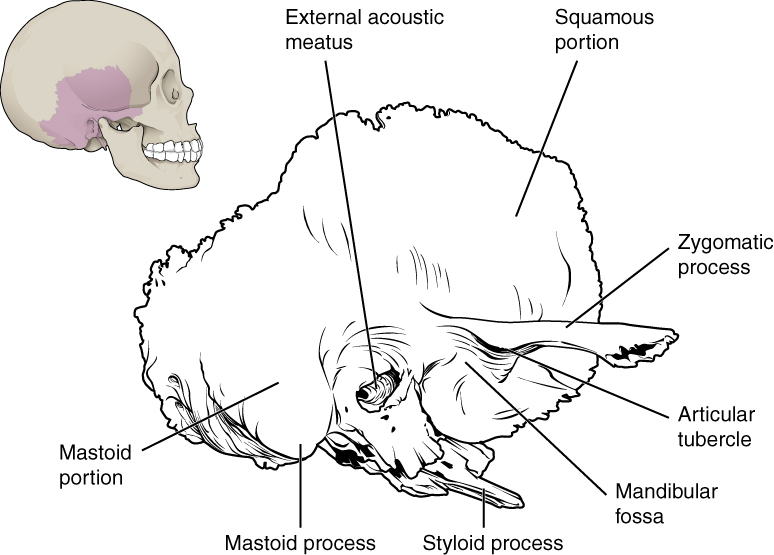
Important landmarks of the temporal bone include the following:
- External acoustic meatus (ear canal)—This is the large opening on the lateral side of the skull that is associated with the ear.
- Mandibular fossa—This is the deep, oval-shaped depression located on the external base of the skull, just in front of the external acoustic meatus. The mandible (lower jaw) joins with the skull at this site as part of the temporomandibular joint, which allows for movements of the mandible during the opening and closing of the mouth.
- Styloid process—Posterior to the mandibular fossa on the external base of the skull is an elongated, downward bony projection called the styloid process, so named because of its resemblance to a stylus (a pen or writing tool). This structure serves as an attachment site for several small muscles and for a ligament that supports the hyoid bone of the neck.
- Mastoid process – The inferior portion of the mastoid portion is an area for muscle attachment (sternocleidomastoid muscle).
- Stylomastoid foramen—This small opening is located between the styloid process and mastoid process. This is the point of exit for the cranial nerve that supplies the facial muscles.
- Carotid canal—The carotid canal is a zig-zag shaped tunnel that provides passage through the base of the skull for the internal carotid artery.

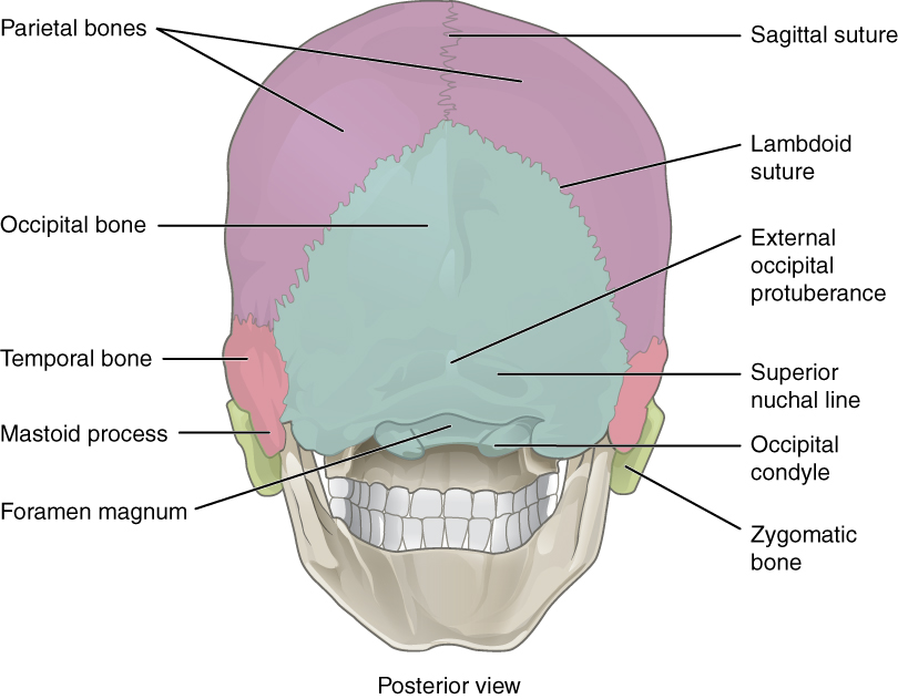
Occipital Bone
The occipital bone is the single bone that forms the posterior skull and posterior base of the cranial cavity. On the base of the skull, the occipital bone contains the large opening of the foramen magnum, which allows for the passage of the spinal cord as it exits the skull. On either side of the foramen magnum is an oval-shaped occipital condyle. These condyles form joints with the first cervical vertebra and thus support the skull on top of the vertebral column.
Sphenoid Bone
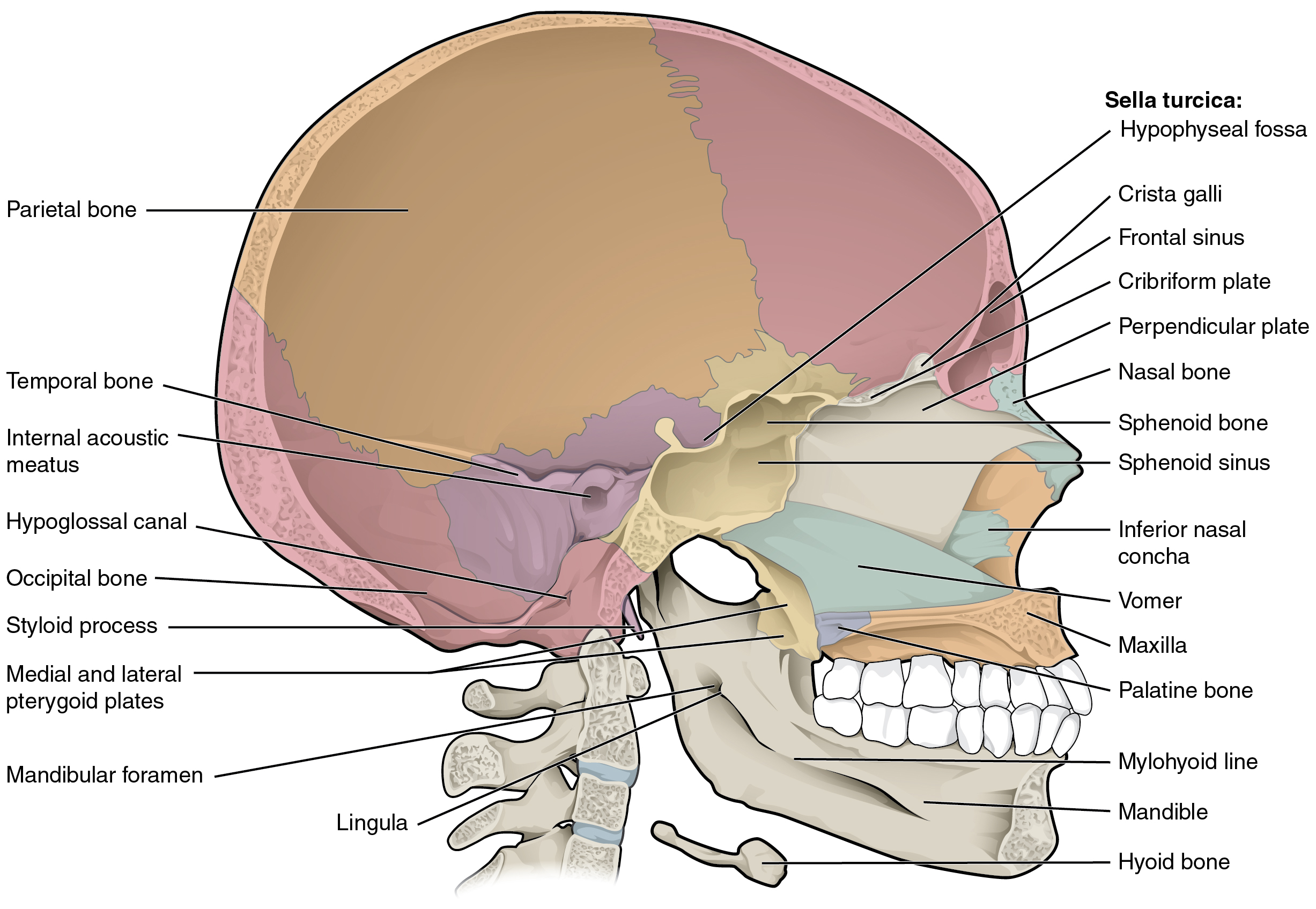
The sphenoid bone is a single, complex bone of the central skull. It serves as a “keystone” bone because it joins with almost every other bone of the skull. The sphenoid forms much of the base of the central skull and also extends laterally to contribute to the sides of the skull. Inside the cranial cavity, the right and left lesser wings of the sphenoid bone, which resemble the wings of a flying bird, form the lip of a prominent ridge that marks the boundary between the anterior and middle cranial fossae. The sella turcica (“Turkish saddle”) is located at the midline of the middle cranial fossa and houses the pituitary gland. The greater wings of the sphenoid bone extend laterally to either side away from the sella turcica, where they form the anterior floor of the middle cranial fossa. The greater wing is best seen on the outside of the lateral skull, where it forms a rectangular area immediately anterior to the squamous portion of the temporal bone. Two major passageways in the sphenoid are the optic canal (passageway for the optic nerve (CN) II) and the superior orbital fissure (passageway for cranial nerves (CN) III, IV, V (part), and VI)
Ethmoid Bone
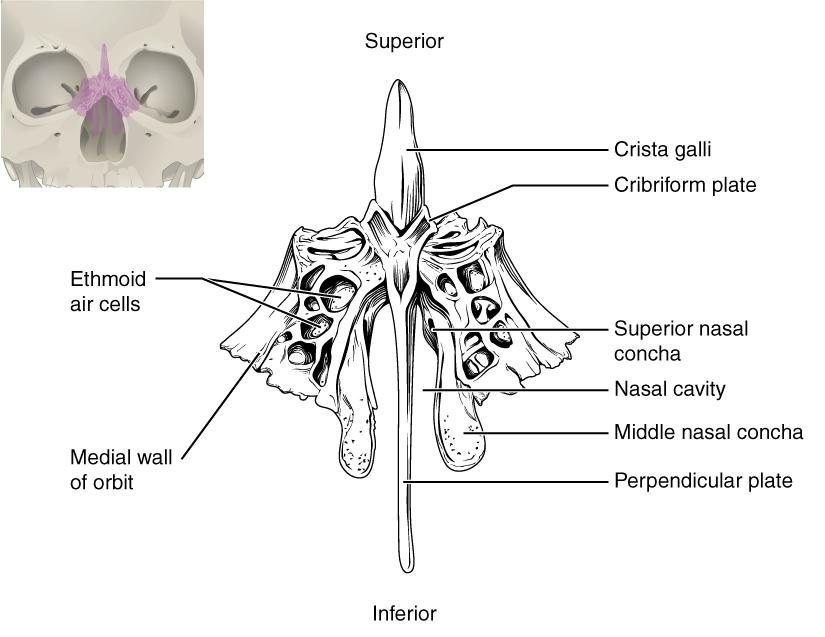
The ethmoid bone is a single, midline bone that forms the roof and lateral walls of the upper nasal cavity, the upper portion of the nasal septum, and contributes to the medial wall of the orbit. On the interior of the skull, the ethmoid also forms a portion of the floor of the anterior cranial cavity.
In the cranial cavity, the ethmoid bone forms a small area at the midline in the floor of the anterior cranial fossa. This region also forms the narrow roof of the underlying nasal cavity. This portion of the ethmoid bone consists of two parts, the crista galli and cribriform plates. The crista galli (“rooster’s comb or crest”) is a small upward bony projection located at the midline. It functions as an anterior attachment point for one of the covering layers of the brain. To either side of the crista galli is the cribriform plate (cribrum = “sieve”), a small, flattened area with numerous small openings called cribriform foramina. Small nerve branches from the olfactory nerves pass through these openings to enter the brain. Located inside the lateral portion of the ethmoid bone are several small, air-filled spaces that are part of the paranasal sinus system of the skull.
Sutures of the Skull
A suture is an immobile joint between adjacent bones of the skull. The narrow gap between the bones is filled with dense, fibrous connective tissue that unites the bones. The long sutures located between the bones of the cranium are not straight, but instead follow irregular, tightly twisting paths. These twisting lines serve to tightly interlock the adjacent bones, thus adding strength to the skull for brain protection.
The two suture lines seen on the top of the skull are the coronal and sagittal sutures. The coronal suture runs from side to side across the skull, within the coronal plane of section. It joins the frontal bone to the right and left parietal bones. The sagittal suture extends posteriorly from the coronal suture, running along the midline at the top of the skull in the sagittal plane of section. It unites the right and left parietal bones. On the posterior skull, the sagittal suture terminates by joining the lambdoid suture. The lambdoid suture extends downward and laterally to either side away from its junction with the sagittal suture. The lambdoid suture joins the occipital bone to the right and left parietal and temporal bones. This suture is named for its upside-down “V” shape, which resembles the capital letter version of the Greek letter lambda (Λ). The squamous suture is located on the lateral skull. It unites the squamous portion of the temporal bone with the parietal bone. At the intersection of four bones is the pterion, a small, capital-H-shaped suture line region that unites the frontal bone, parietal bone, squamous portion of the temporal bone, and greater wing of the sphenoid bone. It is the weakest part of the skull. The pterion is located approximately two finger-widths above the zygomatic arch and a thumb’s width posterior to the upward portion of the zygomatic bone.
Traumatic Brain Injuries – FYI (For Your Information -> you will not be assessed on this content)
Head and traumatic brain injuries are major causes of immediate death and disability, with bleeding and infections as possible additional complications. According to the Centers for Disease Control and Prevention (2010), approximately 30 percent of all injury-related deaths in the United States are caused by head injuries. The majority of head injuries involve falls. They are most common among young children (ages 0–4 years), adolescents (15–19 years), and the elderly (over 65 years). Additional causes vary, but prominent among these are automobile and motorcycle accidents.
Strong blows to the brain-case portion of the skull can produce fractures. These may result in bleeding inside the skull with subsequent injury to the brain. The most common is a linear skull fracture, in which fracture lines radiate from the point of impact. Other fracture types include a comminuted fracture, in which the bone is broken into several pieces at the point of impact, or a depressed fracture, in which the fractured bone is pushed inward. In a contrecoup (counterblow) fracture, the bone at the point of impact is not broken, but instead a fracture occurs on the opposite side of the skull. Fractures of the occipital bone at the base of the skull can occur in this manner, producing a basilar fracture that can damage the artery that passes through the carotid canal.
A blow to the lateral side of the head may fracture the bones of the pterion. The pterion is an important clinical landmark because located immediately deep to it on the inside of the skull is a major branch of an artery that supplies the skull and covering layers of the brain. A strong blow to this region can fracture the bones around the pterion. If the underlying artery is damaged, bleeding can cause the formation of a hematoma (collection of blood) between the brain and interior of the skull. As blood accumulates, it will put pressure on the brain. Symptoms associated with a hematoma may not be apparent immediately following the injury, but if untreated, blood accumulation will exert increasing pressure on the brain and can result in death within a few hours.
Facial Bones of the Skull
*This content is covered in the pre-lecture assignment and is largely self-study. Parts will be addressed briefly in lecture.
The facial bones of the skull form the upper and lower jaws, the nose, nasal cavity and nasal septum, and the orbit. The facial bones include 14 bones, with six paired bones and two unpaired bones. The paired bones are the maxilla, palatine, zygomatic, nasal, lacrimal, and inferior nasal conchae bones. The unpaired bones are the vomer and mandible bones. Although classified with the brain-case bones, the ethmoid bone also contributes to the nasal septum and the walls of the nasal cavity and orbit.
Maxillary Bone
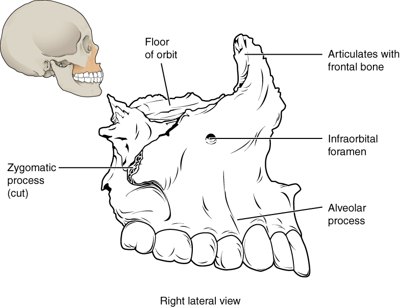
The maxillary bone, often referred to simply as the maxilla (plural = maxillae), is one of a pair that together form the upper jaw, much of the hard palate, the medial floor of the orbit, and the lateral base of the nose. The curved, inferior margin of the maxillary bone that forms the upper jaw and contains the upper teeth is the alveolar process of the maxilla. Each tooth is anchored into a deep socket called an alveolus. On the anterior maxilla, just below the orbit, is the infraorbital foramen. This is the point of exit for a sensory nerve that supplies the nose, upper lip, and anterior cheek. On the inferior skull, the palatine process from each maxillary bone can be seen joining together at the midline to form the anterior three-quarters of the hard palate. The hard palate is the bony plate that forms the roof of the mouth and floor of the nasal cavity, separating the oral and nasal cavities.
Palatine Bone
The palatine bone is one of a pair of irregularly shaped bones that contribute small areas to the lateral walls of the nasal cavity and the medial wall of each orbit. The largest region of each of the palatine bones is the horizontal plate. The plates from the right and left palatine bones join together at the midline to form the posterior quarter of the hard palate. Thus, the palatine bones are best seen in an inferior view of the skull and hard palate.
Cleft Lip and Palate – FYI (For Your Information -> you will not be assessed on this content)
During embryonic development, the right and left maxilla bones come together at the midline to form the upper jaw. At the same time, the muscle and skin overlying these bones join together to form the upper lip. Inside the mouth, the palatine processes of the maxilla bones, along with the horizontal plates of the right and left palatine bones, join together to form the hard palate. If an error occurs in these developmental processes, a birth defect of cleft lip or cleft palate may result.
A cleft lip is a common developmental defect that affects approximately 1:1000 births, most of which are male. This defect involves a partial or complete failure of the right and left portions of the upper lip to fuse together, leaving a cleft (gap).
A more severe developmental defect is a cleft palate, which affects the hard palate. The hard palate is the bony structure that separates the nasal cavity from the oral cavity. It is formed during embryonic development by the midline fusion of the horizontal plates from the right and left palatine bones and the palatine processes of the maxilla bones. Cleft palate affects approximately 1:2500 births and is more common in females. It results from a failure of the two halves of the hard palate to completely come together and fuse at the midline, thus leaving a gap between them. This gap allows for communication between the nasal and oral cavities. In severe cases, the bony gap continues into the anterior upper jaw where the alveolar processes of the maxilla bones also do not properly join together above the front teeth. If this occurs, a cleft lip will also be seen. Because of the communication between the oral and nasal cavities, a cleft palate makes it very difficult for an infant to generate the suckling needed for nursing, thus leaving the infant at risk for malnutrition. Surgical repair is required to correct cleft palate defects.
Zygomatic Bone
The zygomatic bone is also known as the cheekbone. Each of the paired zygomatic bones form much of the lateral walls of the orbit and the lateral-inferior margins of the anterior orbital opening. The short temporal process of the zygomatic bone projects posteriorly, where it forms the anterior portion of the zygomatic arch.
Nasal Bone
The nasal bone is one of two small bones that articulate (join) with each other to form the bony base (bridge) of the nose. They also support the cartilages that form the lateral walls of the nose. These are the bones that are damaged when the nose is broken.
Lacrimal Bone
Each lacrimal bone is a small, rectangular bone that forms the anterior, medial wall of the orbit. The anterior portion of the lacrimal bone forms a shallow depression called the lacrimal fossa, and extending inferiorly from this is the nasolacrimal canal. The lacrimal fluid (tears of the eye), which serves to maintain the moist surface of the eye, drains at the medial corner of the eye into the nasolacrimal canal. This duct then extends downward to open into the nasal cavity, behind the inferior nasal concha. In the nasal cavity, the lacrimal fluid normally drains posteriorly, but with an increased flow of tears due to crying or eye irritation, some fluid will also drain anteriorly, thus causing a runny nose.
Inferior Nasal Conchae
The right and left inferior nasal conchae form a curved bony plate that projects into the nasal cavity space from the lower lateral wall. The inferior concha is the largest of the nasal conchae and can easily be seen when looking into the anterior opening of the nasal cavity.
Vomer Bone
The unpaired vomer bone, often referred to simply as the vomer, is triangular-shaped and forms the posterior-inferior part of the nasal septum. The vomer is best seen when looking from behind into the posterior openings of the nasal cavity.
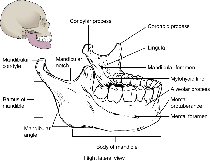
Mandible
The mandible forms the lower jaw and is the only moveable bone of the skull. The side of the mandible consists of a horizontal body and posteriorly, a vertically oriented ramus of the mandible (ramus = “branch”). The outside margin of the mandible, where the body and ramus come together is called the angle of the mandible.
The ramus on each side of the mandible has two upward-going bony projections. The more anterior projection is the flattened coronoid process of the mandible, which provides attachment for one of the biting muscles. The posterior projection is the condylar process of the mandible, which is topped by the oval-shaped condyle. The condyle of the mandible articulates (joins) with the mandibular fossa and articular tubercle of the temporal bone. Together these articulations form the temporomandibular joint, which allows for the opening and closing of the mouth.
Important landmarks for the mandible include the following:
- Alveolar process of the mandible—This is the upper border of the mandibular body and serves to anchor the lower teeth.
- Coronoid process – this protrusion serves as a site for muscular attachment.
- Mandibular condyle – this projection will articulate with the maxilla to form the temporomandibular joint (TMJ).
- Mental foramen—The opening located on each side of the anterior-lateral mandible, which is the exit site for a sensory nerve that supplies the chin.
- Mandibular foramen—This opening is located on the medial side of the ramus of the mandible. The opening leads into a tunnel that runs down the length of the mandibular body. The sensory nerve and blood vessels that supply the lower teeth enter the mandibular foramen and then follow this tunnel. Thus, to numb the lower teeth prior to dental work, the dentist must inject anesthesia into the lateral wall of the oral cavity at a point prior to where this sensory nerve enters the mandibular foramen.
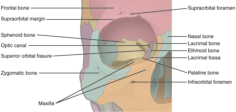
The Orbit
*This content is introduced in the pre-lecture assignment and will be elaborated on in lecture.
The orbit is the bony socket that houses the eyeball and contains the muscles that move the eyeball or open the upper eyelid. Each orbit is cone-shaped, with a narrow posterior region that widens toward the large anterior opening. To help protect the eye, the bony margins of the anterior opening are thickened and somewhat constricted.
The walls of each orbit include contributions from seven skull bones. The frontal bone forms the roof and the zygomatic bone forms the lateral wall and lateral floor. The medial floor is primarily formed by the maxilla, with a small contribution from the palatine bone. The ethmoid bone and lacrimal bone make up much of the medial wall and the sphenoid bone forms the posterior orbit.
At the posterior apex of the orbit is the opening of the optic canal, which allows for passage of the optic nerve from the retina to the brain. Lateral to this is the elongated and irregularly shaped superior orbital fissure, which provides passage for the artery that supplies the eyeball, sensory nerves, and the nerves that supply the muscles involved in eye movements.
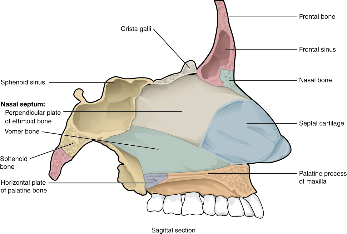
The Nasal Septum and Nasal Conchae
The nasal septum consists of both bone and cartilage components. The upper portion of the septum is formed by the perpendicular plate of the ethmoid bone. The lower and posterior parts of the septum are formed by the triangular-shaped vomer bone. The anterior nasal septum is formed by the septal cartilage, a flexible plate that fills in the gap between the perpendicular plate of the ethmoid and vomer bones. This cartilage also extends outward into the nose where it separates the right and left nostrils. The septal cartilage is not found in the dry skull.
Attached to the lateral wall on each side of the nasal cavity are the superior, middle, and inferior nasal conchae (singular = concha), which are named for their positions. These are bony plates that curve downward as they project into the space of the nasal cavity. They serve to swirl the incoming air, which helps to warm and moisturize it before the air moves into the delicate air sacs of the lungs. This also allows mucus, secreted by the tissue lining the nasal cavity, to trap incoming dust, pollen, bacteria, and viruses. The largest of the conchae is the inferior nasal concha, which is an independent bone of the skull. The middle concha and the superior conchae, which is the smallest, are both formed by the ethmoid bone.
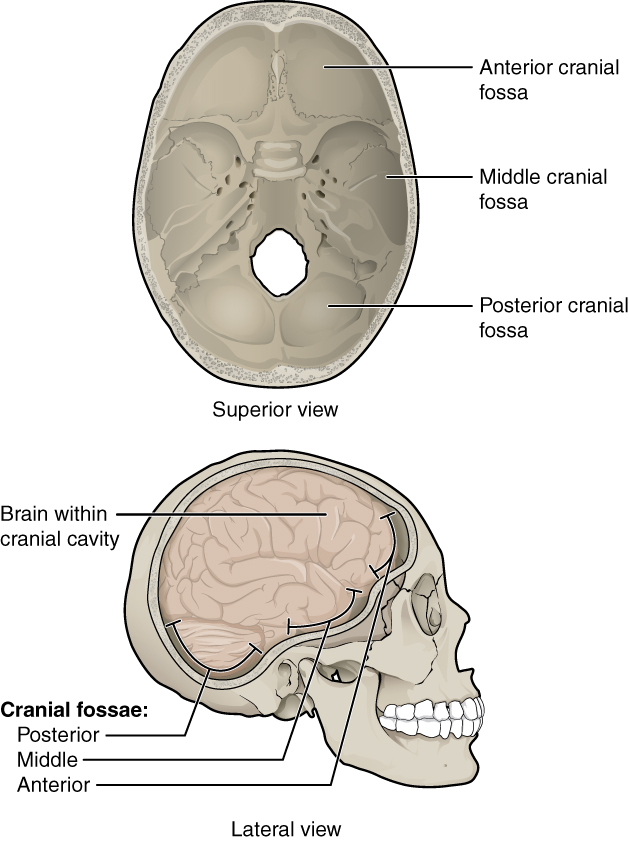
Cranial Fossae
*This content is introduced in the pre-lecture assignment and will be elaborated on in lecture.
Inside the skull, the floor of the cranial cavity is subdivided into three cranial fossae (spaces), which increase in depth from anterior to posterior. Since the brain occupies these areas, the shape of each conforms to the shape of the brain regions that it contains. Each cranial fossa has anterior and posterior boundaries and is divided at the midline into right and left areas by a significant bony structure or opening.
Anterior Cranial Fossa
The anterior cranial fossa is the most anterior and the shallowest of the three cranial fossae. It overlies the orbits and contains the frontal lobes of the brain. Anteriorly, the anterior fossa is bounded by the frontal bone, which also forms the majority of the floor for this space. The lesser wings of the sphenoid bone form the prominent ledge that marks the boundary between the anterior and middle cranial fossae.
Middle Cranial Fossa
The middle cranial fossa is deeper and situated posterior to the anterior fossa. It extends from the lesser wings of the sphenoid bone anteriorly, to the petrous ridges (petrous portion of the temporal bones) posteriorly. The temporal lobes of the brain occupy this fossa. The middle cranial fossa has several openings for the passage of blood vessels and cranial nerves.
Openings in the middle cranial fossa are as follows:
- Optic canal—This opening is located at the anterior lateral corner of the sella turcica. It provides for passage of the optic nerve into the orbit.
- Superior orbital fissure—This large, irregular opening into the posterior orbit is located on the anterior wall of the middle cranial fossa, lateral to the optic canal and under the projecting margin of the lesser wing of the sphenoid bone. Nerves to the eyeball and associated muscles, and sensory nerves to the forehead pass through this opening.
- Foramen rotundum—This rounded opening (rotundum = “round”) is located in the floor of the middle cranial fossa, just inferior to the superior orbital fissure. It is the exit point for a major sensory nerve that supplies the cheek, nose, and upper teeth.
- Foramen ovale of the middle cranial fossa—This large, oval-shaped opening in the floor of the middle cranial fossa provides passage for a major sensory nerve to the lateral head, cheek, chin, and lower teeth.
- Foramen spinosum—This small opening, located posterior-lateral to the foramen ovale, is the entry point for an important artery that supplies the covering layers surrounding the brain. The branching pattern of this artery forms readily visible grooves on the internal surface of the skull and these grooves can be traced back to their origin at the foramen spinosum.
- Carotid canal—This is the zig-zag passageway through which a major artery to the brain enters the skull. The entrance to the carotid canal is located on the inferior aspect of the skull, anteromedial to the styloid process. From here, the canal runs anteromedially within the bony base of the skull. Just above the foramen lacerum, the carotid canal opens into the middle cranial cavity, near the posterior-lateral base of the sella turcica.
Posterior Cranial Fossa
The posterior cranial fossa is the most posterior and deepest portion of the cranial cavity. It contains the cerebellum of the brain. The posterior fossa is bounded anteriorly by the petrous ridges, while the occipital bone forms the floor and posterior wall. It is divided at the midline by the large foramen magnum (“great aperture”), the opening that provides for passage of the spinal cord.
Located on the medial wall of the petrous ridge in the posterior cranial fossa is the internal acoustic meatus. This opening provides for passage of the nerve from the hearing and equilibrium organs of the inner ear, and the nerve that supplies the muscles of the face. Located at the anterior-lateral margin of the foramen magnum is the hypoglossal canal. These emerge on the inferior aspect of the skull at the base of the occipital condyle and provide passage for an important nerve to the tongue.
Immediately inferior to the internal acoustic meatus is the large, irregularly shaped jugular foramen. Several cranial nerves from the brain exit the skull via this opening. It is also the exit point through the base of the skull for all the venous return blood leaving the brain. The venous structures that carry blood inside the skull form large, curved grooves on the inner walls of the posterior cranial fossa, which terminate at each jugular foramen.
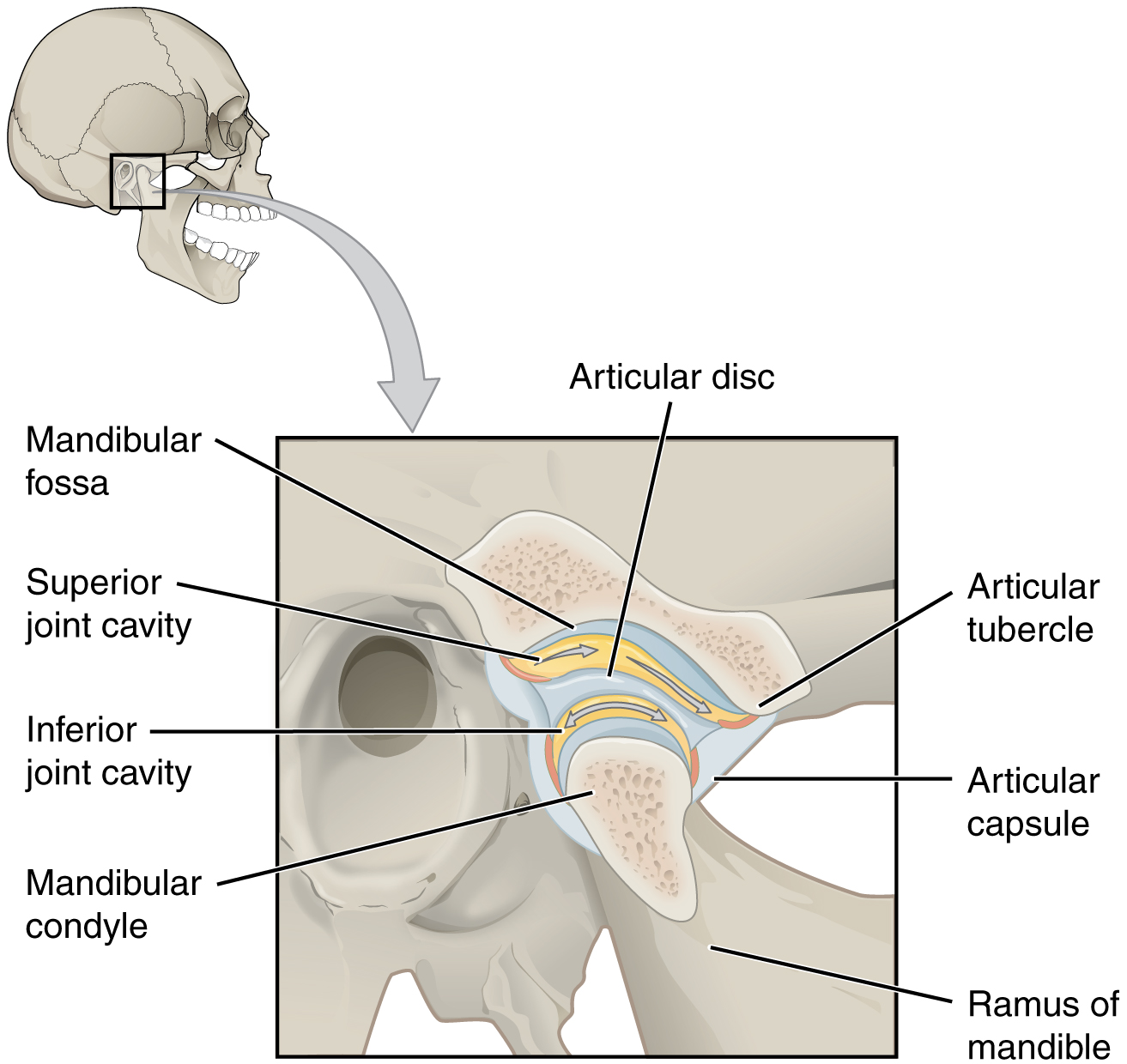
Temporomandibular Joint
The temporomandibular joint (TMJ) is the joint that allows for opening (mandibular depression) and closing (mandibular elevation) of the mouth, as well as side-to-side and protraction/retraction motions of the lower jaw. This joint involves the articulation between the mandibular fossa and articular tubercle of the temporal bone, with the condyle (head) of the mandible. Located between these bony structures, filling the gap between the skull and mandible, is a flexible articular disc. This disc serves to smooth the movements between the temporal bone and mandibular condyle.
TMJ – Fun Facts (-> you will not be assessed on this content)
Movement at the TMJ during opening and closing of the mouth involves both gliding and hinge motions of the mandible. With the mouth closed, the mandibular condyle and articular disc are located within the mandibular fossa of the temporal bone. During the opening of the mouth, the mandible hinges downward and at the same time is pulled anteriorly, causing both the condyle and the articular disc to glide forward from the mandibular fossa onto the downward projecting articular tubercle. The net result is a forward and downward motion of the condyle and mandibular depression. The temporomandibular joint is supported by an extrinsic ligament that anchors the mandible to the skull. This ligament spans the distance between the base of the skull and the lingula on the medial side of the mandibular ramus.
Dislocation of the TMJ may occur when opening the mouth too wide (such as when taking a large bite) or following a blow to the jaw, resulting in the mandibular condyle moving beyond (anterior to) the articular tubercle. In this case, the individual would not be able to close his or her mouth. Temporomandibular joint disorder is a painful condition that may arise due to arthritis, wearing of the articular cartilage covering the bony surfaces of the joint, muscle fatigue from overuse or grinding of the teeth, damage to the articular disc within the joint, or jaw injury. Temporomandibular joint disorders can also cause headache, difficulty chewing, or even the inability to move the jaw (lock jaw). Pharmacologic agents for pain or other therapies, including bite guards, are used as treatments.
Muscles That Create Facial Expression
*This content is introduced in the pre-lecture assignment and will be elaborated on in lecture.
The origins of the muscles of facial expression are on the surface of the skull. The insertions of these muscles have fibers intertwined with connective tissue and the dermis of the skin. Because the muscles insert in the skin rather than on bone, when they contract, the skin moves to create facial expression.
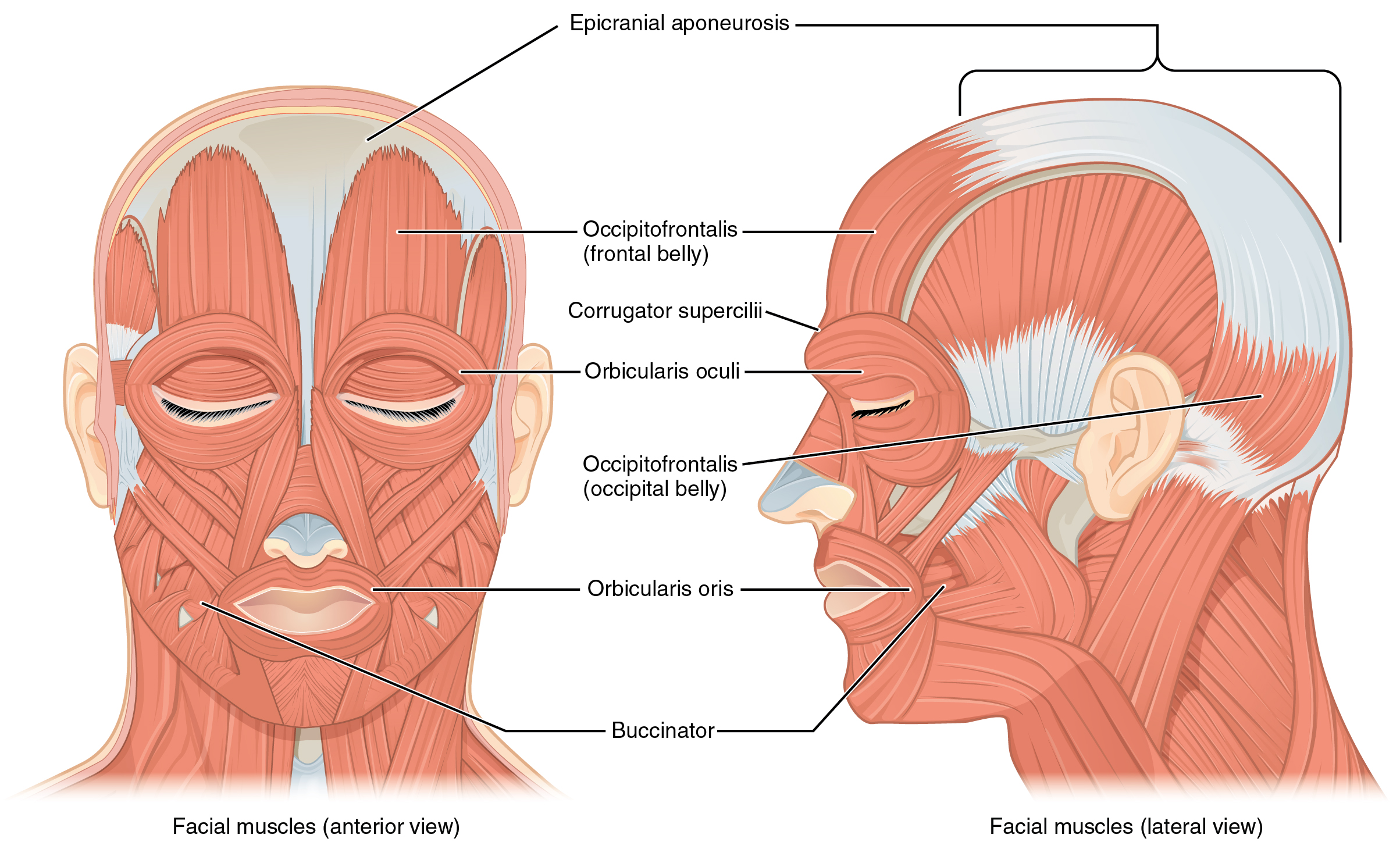
The muscles of facial expression are discussed further in the lecture videos. Focus on the action created by the muscles of facial expression and what structures the muscles act upon.
| Muscle | Action | Target | Innervation |
| Occipitofrontalis – Frontal Belly | Elevate eyebrows, wrinkle forehead | Scalp | CN VII |
| Occiptofrontalis – Occipital Belly | Retract scalp | Scalp | CN VII |
| Corrugator Supercilii | Pull eyebrow inferior and medial, wrinkle superior nose (vertically) | Scalp/Eye | CN VII |
| Orbicularis Oculi | Close eyelids | Eye | CN VII |
| Levator Palpebrae Superioris | Elevate superior eyelid | Eye | CN III** |
| Procerus | Depress medial eyebrow, wrinkle superior nose (horizontally) | Nose | CN VII |
| Nasalis | Depress the nasal cartilage | Nose | CN VII |
| Levator Labii Superioris Alaeque Nasi | Elevate upper lip, dilates nostrils | Nose/Mouth | CN VII |
| Levator Anguli Oris | Widens mouth, elevates corner of mouth | Mouth | CN VII |
| Levator Labii Superioris | Elevates and furrows upper lip | Mouth | CN VII |
| Buccinator | Compresses cheek, draws corner of mouth laterally | Mouth | CN VII |
| Risorius | Draws corner of lip laterally | Mouth | CN VII |
| Orbicularis Oris | Closes lips, purses, and protrudes lips | Mouth | CN VII |
| Zygomaticus Major | Raises lateral corner of mouth | Mouth | CN VII |
| Zygomaticus Minor | Raises lateral corner of mouth | Mouth | CN VII |
| Depressor Angui Oris | Draws the corner of mouth inferiorly and laterally | Mouth | CN VII |
| Depressor Labii Inferioris | Depresses lower lip | Mouth | CN VII |
| Mentalis | Wrinkles skin of chin, protrudes lower lip | Mouth | CN VII |
| Platysma | Tenses skin of inferior face and neck | Neck | CN VII |
