
Module 9: Human Development and Anatomy Through the Lifespan
Learning Objectives:
By the end of this class, students will be able to:
- Identify key times and events in embryological development and describe the consequences of teratogen interference during those times.
- Understand critical periods of development and some of the consequences of disrupting typical development.
- Describe common changes that occur in the body with aging.
Terms to Know
|
*Covered only in lecture, not in this text |
The topic of embryonic and fetal development could fill an entire course. This will only be a brief introduction to the topic.
Embryonic and fetal ages are expressed in terms of weeks from fertilization. The period of time required for full development of a fetus in utero is referred to as gestation. It can be subdivided into distinct periods:
- Embryonic period: from fertilization to week 8.
- Fetal period: from week nine until birth.
Embryonic Development
This information will be covered in the assignment and reviewed and built upon in lecture.
The first two weeks of prenatal development are sometimes referred to as the pre-embryonic stage. By the third week, the tissues that ultimately develop into all organ systems of the body have formed. By the end of the embryonic period, all of the organ systems are structured in rudimentary form, although the organs themselves are either nonfunctional or only semi-functional. The process of organ formation is called organogenesis.
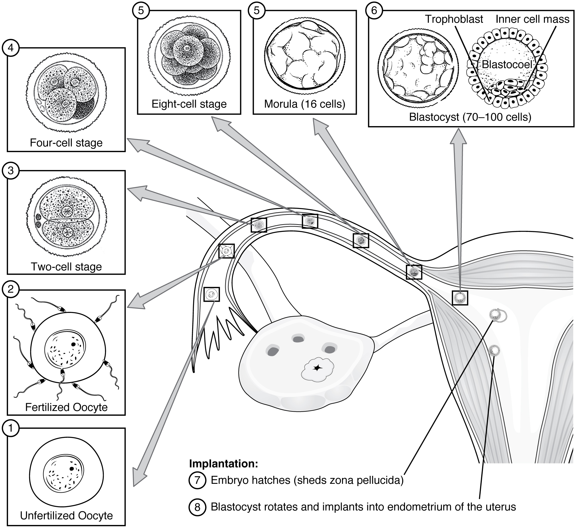
Week 1 (Pre-Embryonic Period)
The terminology “embryo” is not typically used this early in gestation, but we will not ask you to know the other terms, so we will stick with “embryo” for simplicity.
Following fertilization, during the journey to the uterus, repeated rapid cell division occurs. Approximately 3 days after fertilization, a 16-cell embryo reaches the uterus. Once inside the uterus, the embryo floats freely for several more days, continuing to divide. At the end of the first week, the embryo embeds itself in the uterine lining, beginning the implantation process.
Week 2 (Pre-Embryonic Period)
At the beginning of the second week, the cells form into a two-layered disc of embryonic cells. Little embryonic growth occurs at this stage. Instead, the second week is a time when structures that support the embryo form. The amniotic fluid forms, which allows the embryo to float within it, protecting it from trauma and rapid temperature changes. The placenta begins to form in this stage as well. This is critically important because the embryo and fetus need oxygen, nutrients, and other factors to develop and grow. The placenta begins to form very early after implantation in order to provide these critical resources and support the development of all organ systems. Though it takes a few weeks before the placenta takes over this entire role, its development starts very early in gestation.
Week 3
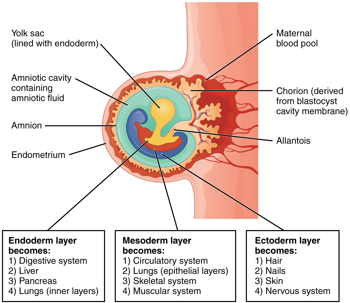
As the third week of development begins, the embryo takes the form of a three-layered oval-shaped disc. This disc has caudal and cranial ends. Caudal means “tail” and cranial means “head,” so even though it doesn’t look like it yet, the head end and tail end of the embryo is already established.
Each layer of the three-layer disc will develop into specific structures in the embryo. You don’t need to know the specific structures that come from each layer, but we want you to understand that all tissues of the fully-developed human body originate from these three tissue layers, and specific layers develop specific structures.
Organ formation begins during the third week. The human heart is the first functional organ to develop. It forms around 18 to 19 days after fertilization. The heart begins beating and pumping blood around day 21 or 22, a mere three weeks after fertilization. This emphasizes the critical nature of the heart in distributing blood through the vessels and the vital exchange of nutrients, oxygen, and wastes both to and from the developing baby.
The nervous system also begins to form in the third week. Adequate folic acid is critical for nervous system development.
Week 4
Morphogenesis, or a change in shape, occurs in the fourth week. The embryo, which begins as a flat sheet of cells, begins to acquire a cylindrical shape through the process of embryonic folding. The embryo folds laterally (transverse folding) and again at either end (cephalocaudal folding), forming a C-shape with head and tail ends that are now distinct. The transverse folding essentially creates a tube, called the primitive gut.
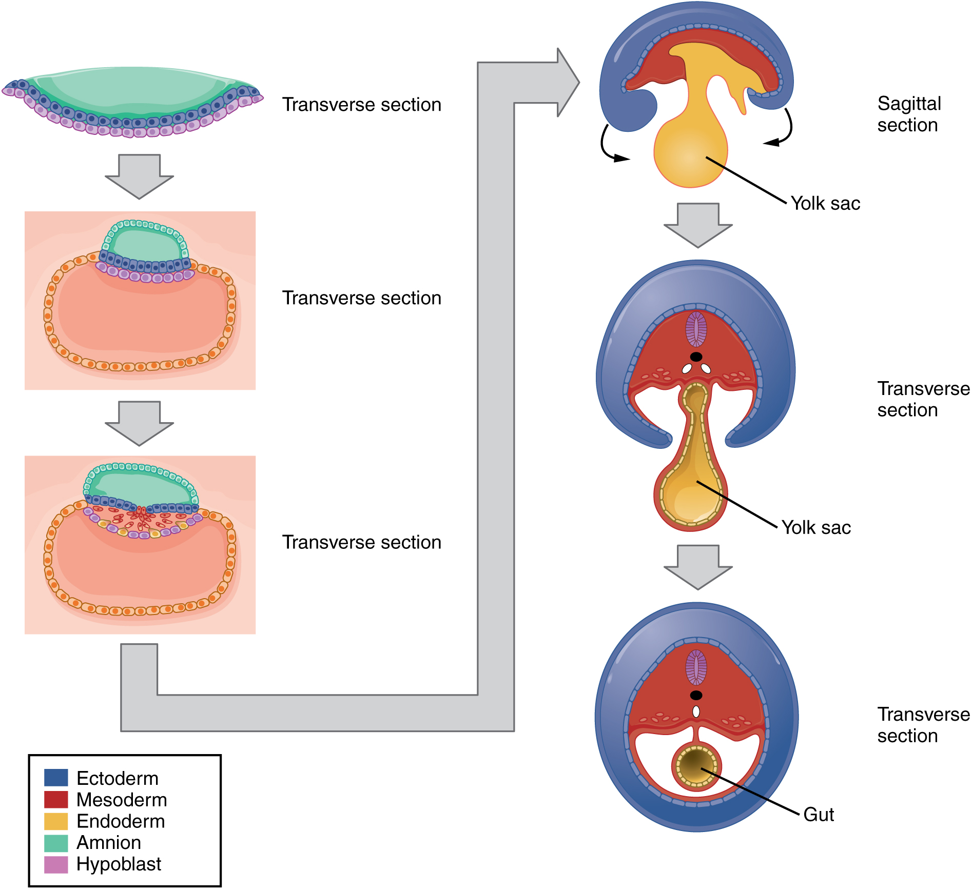
By the end of the fourth week, all organ systems exist and are in the process of forming, though they have a long way to go before they are even considered rudimentary structures.
Limb Development in Weeks 4-8
During weeks 4–5 the limbs begin to form. The upper limbs start to form a few days before the lower limbs. They both begin as limb buds emerging from the embryo body wall. By the 7th week, muscles are capable of contraction, and uncontrolled fetal limb movements begin to occur. The hands and feet are initially webbed, but they develop separate fingers and toes in weeks 7 and 8.
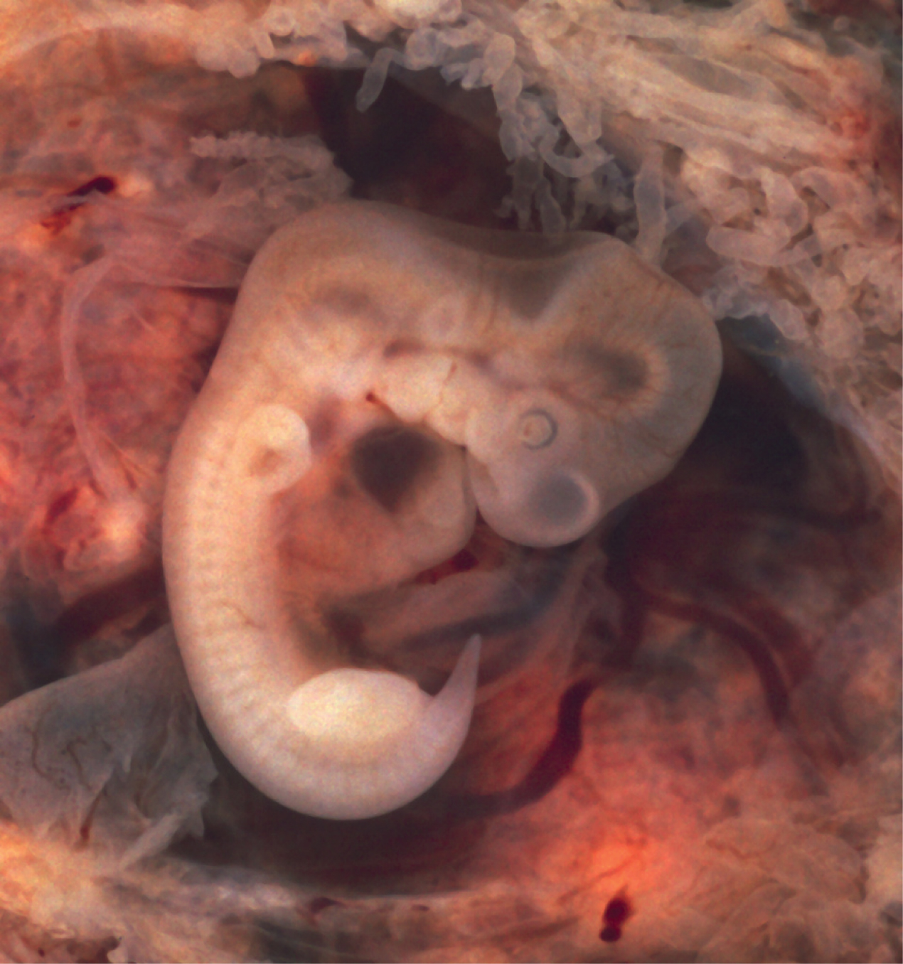
Completing the Embryonic Period
By the eighth week, the head is nearly as large as the rest of the embryo’s body, and all major brain structures are in place. The external genitalia are apparent, but at this point, male and female embryos are indistinguishable. Bone begins to replace cartilage in the embryonic skeleton through the process of ossification. By the end of the embryonic period, all of the organ systems are present in their rudimentary form. The embryo is approximately 3 cm (1.2 in).
Fetal Development
A developing human is called a fetus from the ninth week of gestation until birth. This 30-week period of development is marked by continued cell growth and differentiation, which fully develop the structures and functions of the immature organ systems formed during the embryonic period. The completion of fetal development results in a newborn who, although still immature in many ways, is capable of survival outside the womb.
We do not expect you to know the details of fetal period development. However, here are a few highlights, just for fun:
- During weeks 9–12: The brain continues to expand and the body elongates. The bone marrow begins to take over the process of erythrocyte production—a task that the liver performed during the embryonic period. The liver now secretes bile. The eyes are well-developed by this stage, but the eyelids are fused shut. The fingers and toes begin to develop nails.
- Weeks 13–16: Blinking motions begin, although the eyes remain sealed shut. The lips exhibit sucking motions. The scalp begins to grow hair.
- Weeks 16–20: The fetus grows and limb movements become more powerful, the mother may begin to feel quickening, or fetal movements. Sebaceous glands coat the skin with a waxy, protective substance that protects and moisturizes the skin and may provide lubrication during childbirth.
- Weeks 21–30: Characterized by rapid weight gain. Axons of the spinal cord begin to be myelinated. (The process of myelination is not completed until into adulthood.) During this period, the fetus grows eyelashes. The eyelids are no longer fused and can be opened and closed. The lungs begin producing surfactant, a substance that reduces surface tension in the lungs and assists proper lung expansion after birth. By week 28, enough alveoli have matured that a baby born prematurely at this time can usually breathe on its own, though alveoli continue to form until adulthood. In male fetuses, the testes descend into the scrotum near the end of this period.
- Weeks 31-Birth: The fetus continues to grow, develop alveoli, and lay down fat for insulation after birth.
Anatomy through the lifespan
This section will be covered in lecture, but there are a few main takeaways we’ll mention here.
First, whether for good or bad, what we do when we are young can affect our anatomy when we are older. We can do weight bearing exercise to build bone density and delay bone loss as we age. We can continue to exercise our brain to delay the effects of aging or neurodegenerative disease (if present). In contrast, smoking or significant sun exposure can both increase the risk of cancer later in life.
Second, when an exposure or incident occurs, or in some cases doesn’t occur, during development it matters. Critical periods of development are times when development is occurring very rapidly in a given structure. They also represent windows of vulnerability, when an incident or exposure that disrupts that development even a small amount can have greater consequences than during other times in development. For example, lead poisoning has greater neurological consequences when it occurs in young children than when adults are exposed.
Consequences of Aging
This information will be covered in lecture.
The outward signs of aging are easily recognizable. The skin and other tissues become thinner and drier, reducing their elasticity, contributing to wrinkles and high blood pressure. The face looks flabby because elastic and collagen fibers decrease in connective tissue. Glasses and hearing aids may become parts of life as the senses slowly deteriorate, all due to reduced elasticity. Cells are less able to divide and regenerate. Wound healing is slower in the elderly, accompanied by a higher frequency of infection as the capacity of the immune system to fend off pathogen declines.
Overall height decreases as the bones lose calcium and other minerals. With age, fluid decreases in the fibrous cartilage disks intercalated between the vertebrae in the spine. Joints lose cartilage and stiffen. Many tissues, including muscle tissue, lose mass through a process called atrophy. Age-related muscle atrophy is called sarcopenia.
The brain and spinal cord lose neurons. Nerves do not transmit impulses with the same speed and frequency as in the past. Some loss of thought clarity and memory can accompany aging.
These are just a few of the consequences of aging. We will discuss the impact of aging on specific body systems and regions throughout this course.
