Module 35: Lower Extremity III – Leg, Ankle, and Foot
Learning Objectives:
By the end of this class, students will be able to:
- Describe the thigh and leg muscles that act on the knee.
- Describe the bones and joints of the ankle and foot.
- Describe the muscles of the leg and foot.
- Group the muscles of the leg and foot by their location and/or function.
- Review the anatomy of the lower extremity using clinical application, grouping, and other review methods.
Terms to Know
|
Bones of the Lower Extremity
Joints of the Lower Extremity
Other Lower Extremity Terms
|
Muscles of the Lower Extremity
*Covered only in lecture, not in this text |
Tibia
This content will be covered in the assignment and briefly reviewed in lecture.
The shaft of the tibia is triangular in shape. Both the anterior tibial border and the medial side of the triangular shaft are located immediately under the skin and can be easily palpated along the entire length of the tibia. The interosseous membrane of the leg is the sheet of dense connective tissue that holds the tibia and fibula bones together.
The large expansion found on the medial side of the distal tibia is the medial malleolus. This forms the large bony bump found on the medial side of the ankle region. Both the smooth surface on the inside of the medial malleolus and the smooth area at the distal end of the tibia articulate with the talus bone of the foot as part of the ankle joint. The lateral side of the distal tibia articulates with the distal end of the fibula, forming the distal tibiofibular joint.
Fibula
This content will be covered in the assignment and briefly reviewed in lecture.
The medial border of the thin shaft of the fibula attaches to the interosseous membrane. The distal end of the fibula forms the lateral malleolus, which forms the easily palpated bony bump on the lateral side of the ankle. The medial side of the lateral malleolus articulates with the talus of the foot as part of the ankle joint. The distal fibula also articulates with the tibia.
Bones and Joints of the foot
Tarsal Bones
This content will be covered in the assignment and briefly reviewed in lecture.
The posterior half of the foot is formed by seven tarsal bones. The most superior bone is the talus. This has a relatively square-shaped, upper surface that articulates with the tibia and fibula to form the ankle joint. Inferiorly, the talus articulates with the calcaneus, the largest bone of the foot, which forms the heel. Body weight is transferred from the tibia to the talus to the calcaneus, which rests on the ground.
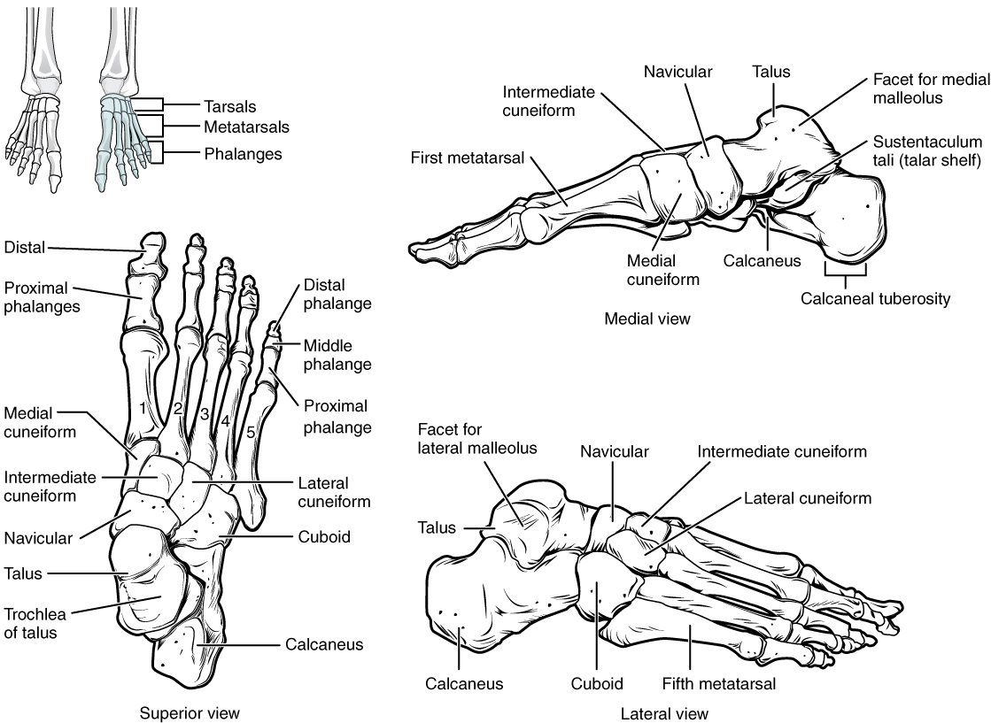
The cuboid articulates with the anterior end of the calcaneus bone. The talus articulates anteriorly with the navicular, which in turn articulates anteriorly with the three cuneiform (“wedge-shaped”) bones. These bones are the medial cuneiform, the intermediate cuneiform, and the lateral cuneiform. Each of these bones has a broad superior surface and a narrow inferior surface, which together produce the transverse (medial-lateral) curvature of the foot. The navicular and lateral cuneiform also articulate with the medial side of the cuboid bone. The joints between the tarsal bones are called the intertarsal joint.
Metatarsal Bones
The anterior half of the foot is formed by the five metatarsal bones, which are located between the tarsal bones of the posterior foot and the phalanges of the toes. These elongated bones are numbered I-V, starting with the medial side of the foot. The first metatarsal bone is shorter and thicker than the others. The second metatarsal is the longest. The base of the metatarsal bone is the proximal end of each metatarsal bone. These articulate with the cuboid or cuneiform bones, forming the tarsometatarsal joints. The base of the fifth metatarsal has a large, lateral expansion that provides a muscle attachment for the fibularis (peroneus) brevis. This expanded base of the fifth metatarsal can be felt as a bony bump at the midpoint along the lateral border of the foot. The expanded distal end of each metatarsal is the head of the metatarsal bone. Each metatarsal bone articulates with the proximal phalanx of a toe to form a metatarsophalangeal joint. The heads of the metatarsal bones also rest on the ground and form the ball (anterior end) of the foot.
Phalanges
The toes contain a total of 14 phalanges (singular phalanx), arranged in a similar manner as the phalanges of the fingers. The toes are numbered 1–5, starting with the big toe, or the hallux. The big toe has two phalanx bones, the proximal and distal phalanges. The remaining toes all have proximal, middle, and distal phalanges. A joint between adjacent phalanx bones is called an interphalangeal joint. The hallux only has an interphalangeal joint, while digits 2-5 have a proximal interphalangeal joint and a distal interphalangeal joint.
Arches of the Foot
This content will be covered in lecture.
When the foot comes into contact with the ground during walking, running, or jumping , the impact of the body weight puts a tremendous amount of pressure and force on the foot. The bones, joints, ligaments, and muscles of the foot absorb this force, thus greatly reducing the amount of shock that is passed superiorly into the lower limb and body. The arches of the foot play an important role in this shock-absorbing ability. When weight is applied to the foot, these arches will flatten somewhat, thus absorbing energy. When the weight is removed, the elastic ligaments maintaining the arches recoil, releasing the stored energy and improving the energy efficiency of walking. Contraction of the foot muscles also plays an important role in this energy absorption. The arches also serve to distribute body weight side to side and to either end of the foot.
The foot has a transverse arch, a medial longitudinal arch, and a lateral longitudinal arch. The transverse arch forms the medial-lateral curvature of the mid-foot. This arch helps to distribute body weight from side to side within the foot, thus allowing the foot to accommodate uneven terrain. The longitudinal arches run down the length of the foot. The lateral longitudinal arch is relatively flat, whereas the medial longitudinal arch is larger (taller).
The principal support for the longitudinal arches of the foot is a deep fascia called plantar aponeurosis, which runs from the calcaneus bone to the toes. The plantar fascia is superficial to the intrinsic muscles of the foot.
Ankle Joint
This content will be covered in the assignment and briefly reviewed in lecture.
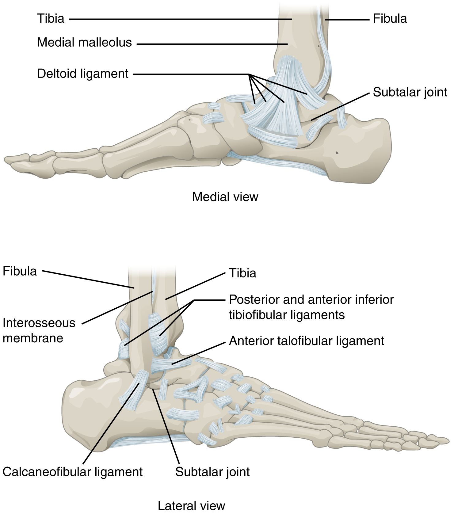
The ankle is formed by the talocrural joint. It consists of the articulations between the talus bone of the foot and the distal ends of the tibia and fibula of the leg (crural = “leg”). The superior aspect of the talus bone is square-shaped and has three areas of articulation. The top of the talus articulates with the inferior tibia. This is the portion of the ankle joint that carries the body weight between the leg and foot. The sides of the talus are firmly held in position by the articulations with the medial malleolus of the tibia and the lateral malleolus of the fibula, which prevent any side-to-side motion of the talus. The ankle is thus a uniaxial hinge joint that allows only for dorsiflexion and plantar flexion of the foot.
Additional joints between the tarsal bones of the posterior foot allow for the movements of foot inversion and eversion. These motions do not happen at the ankle, but at intertarsal joints of the foot.
The talocrural joint of the ankle is supported by several strong ligaments located on the sides of the joint. On the medial side is the broad deltoid ligament. The deltoid ligament supports the ankle joint and also resists excessive eversion of the foot. The lateral side of the ankle has several smaller ligaments. These include the anterior talofibular ligament and the posterior talofibular ligament, both of which span between the talus bone and the lateral malleolus of the fibula, and the calcaneofibular ligament, located between the calcaneus bone and fibula. These ligaments support the ankle and also resist excess inversion of the foot. The anterior and posterior tibiofibular ligaments hold the distal portions of the tibia and fibula together to support the talocrural joint.
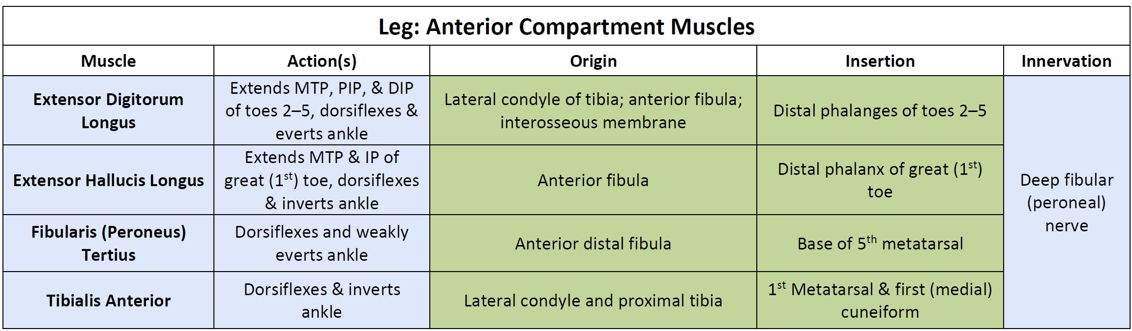
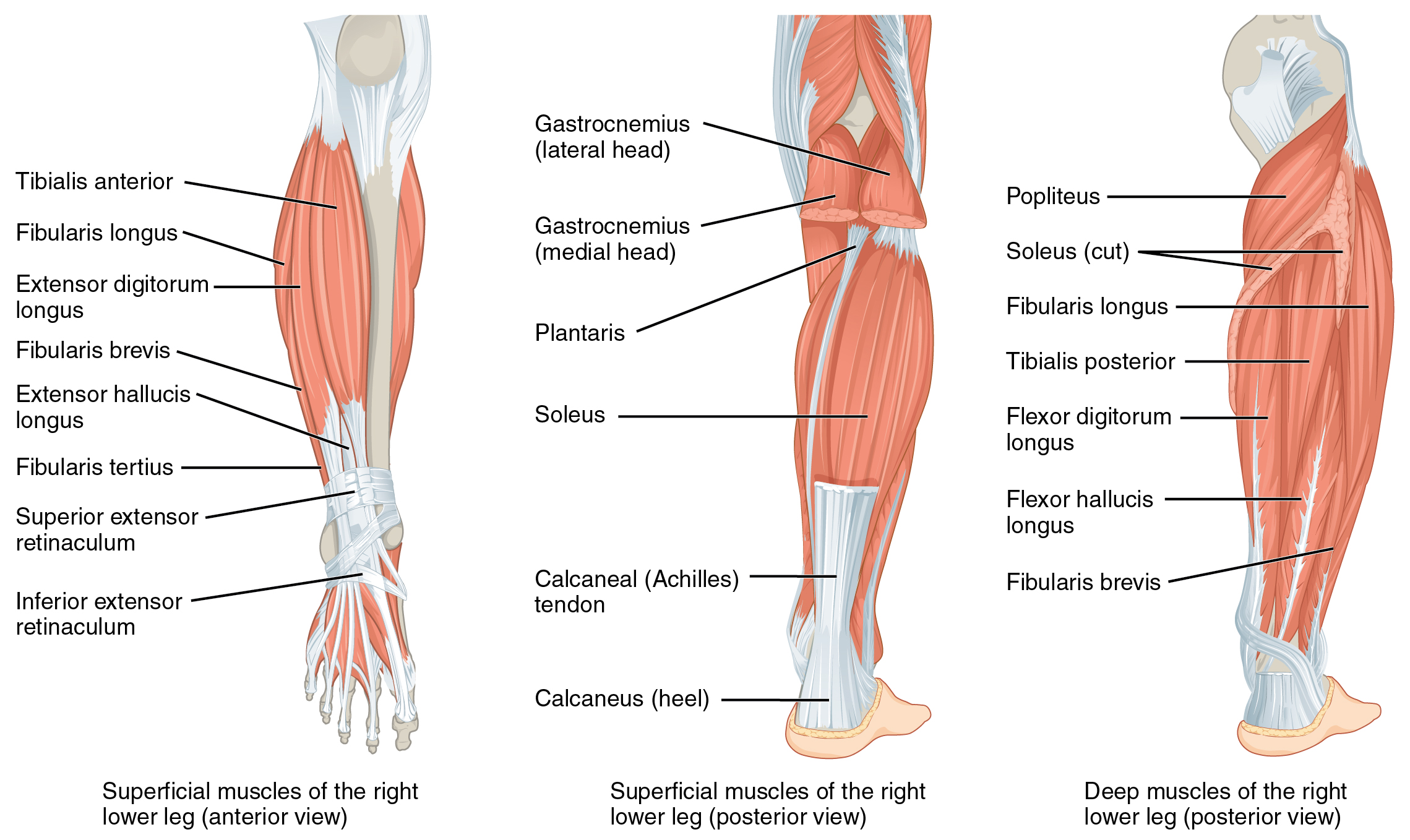
Muscles of the Leg
This content will be covered in the assignment and reviewed in lecture.
The muscles of the leg are divided into four compartments by thick fascia.
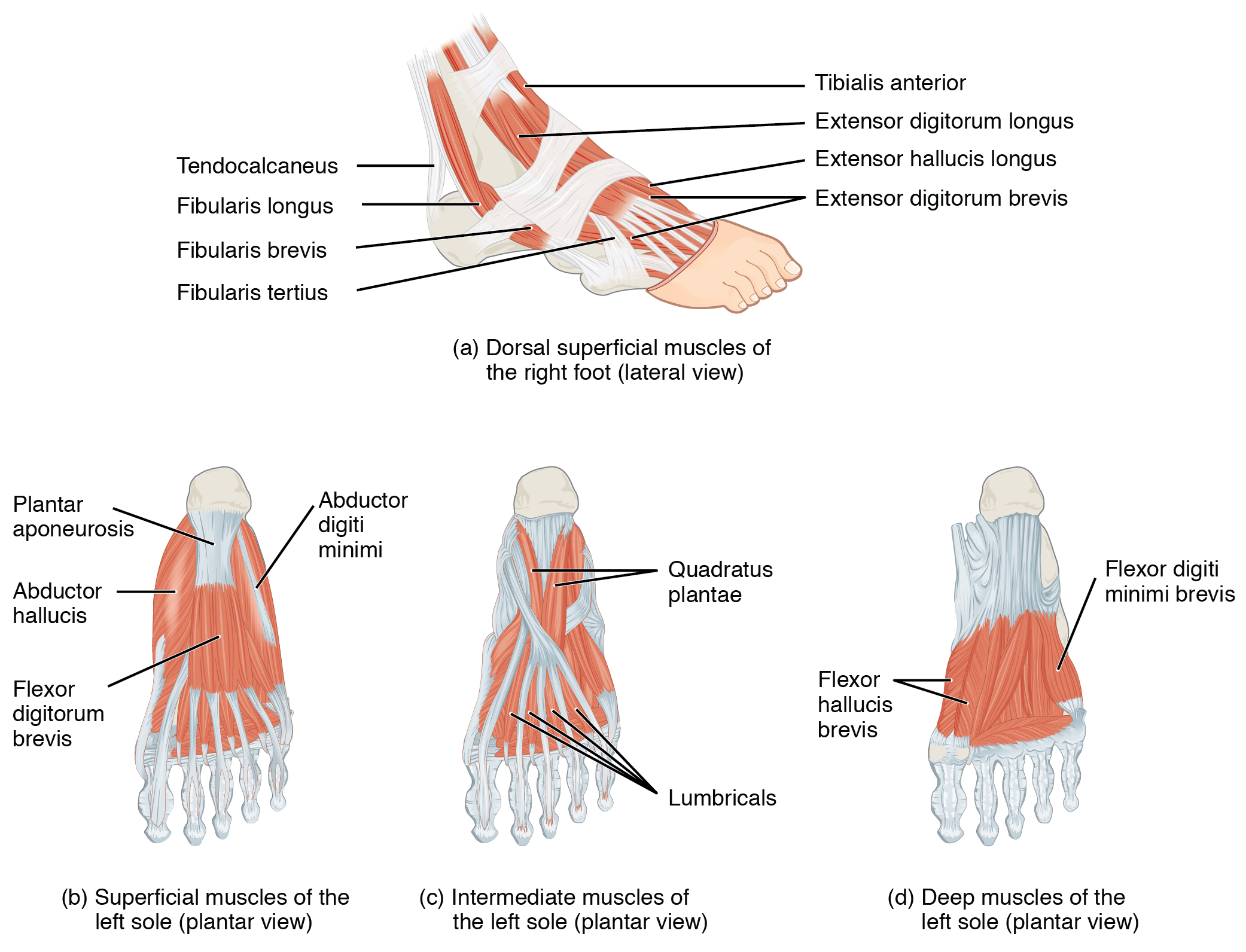
The muscles in the anterior compartment of the leg are the the tibialis anterior, a long and thick muscle on the lateral surface of the tibia, the extensor hallucis longus, deep under it, and the extensor digitorum longus, lateral to it. They all contribute to dorsiflexion. The fibularis tertius, a small muscle that originates on the anterior surface of the fibula, is associated with the extensor digitorum longus and sometimes fused to it, but is not present in all people. Thick bands of connective tissue called the superior extensor retinaculum and inferior extensor retinaculum, hold the tendons of these muscles in place during dorsiflexion.
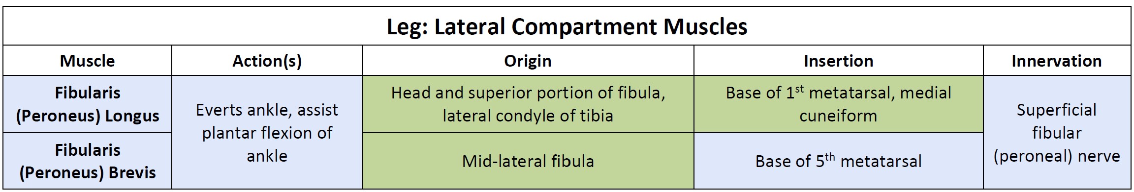
The lateral compartment of the leg includes two muscles: the fibularis longus (peroneus longus) and the fibularis brevis (peroneus brevis).
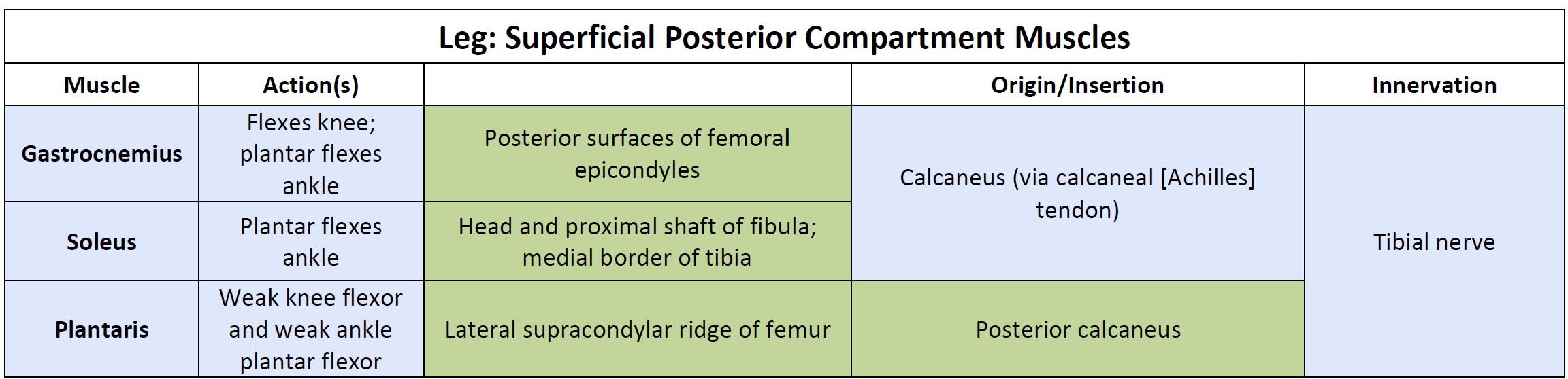
The muscles of the superficial posterior compartment of the leg all insert onto the calcaneus, with two of them forming the calcaneal (Achilles) tendon. The most superficial and visible muscle of the calf is the gastrocnemius. Deep to the gastrocnemius is the wide, flat soleus. The plantaris runs obliquely between the two; some people may have two of these muscles, whereas no plantaris is observed in about seven percent of other cadaver dissections.
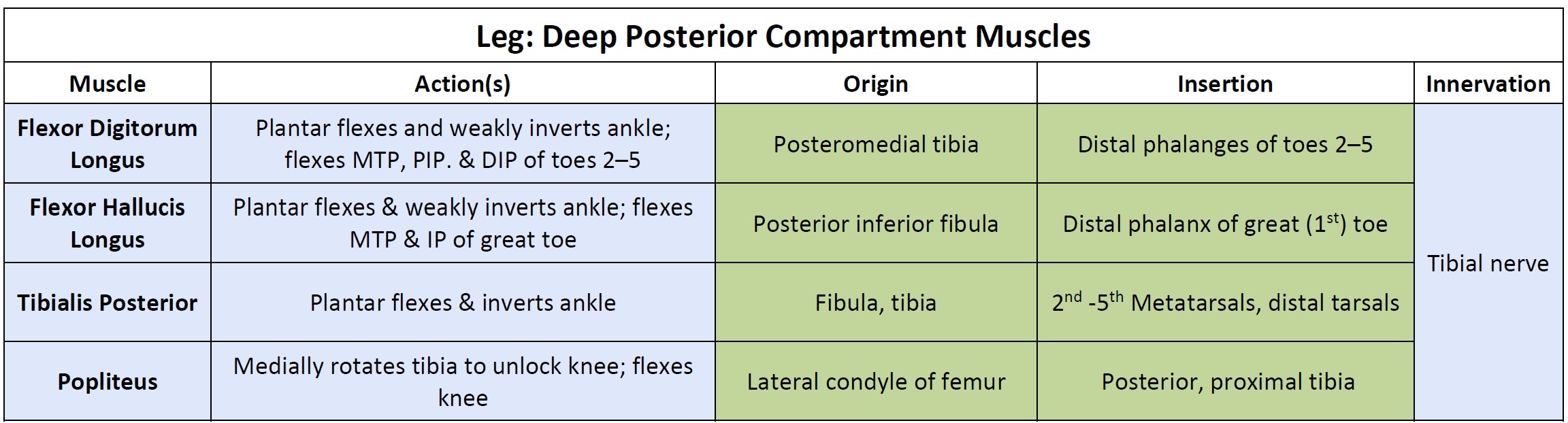
There are four deep muscles in the posterior compartment of the leg as well: the popliteus, flexor digitorum longus, flexor hallucis longus, and tibialis posterior. The popliteus does not cross the ankle joint. Instead it acts on the knee joint to medially rotate and “unlock” the knee from full extension.
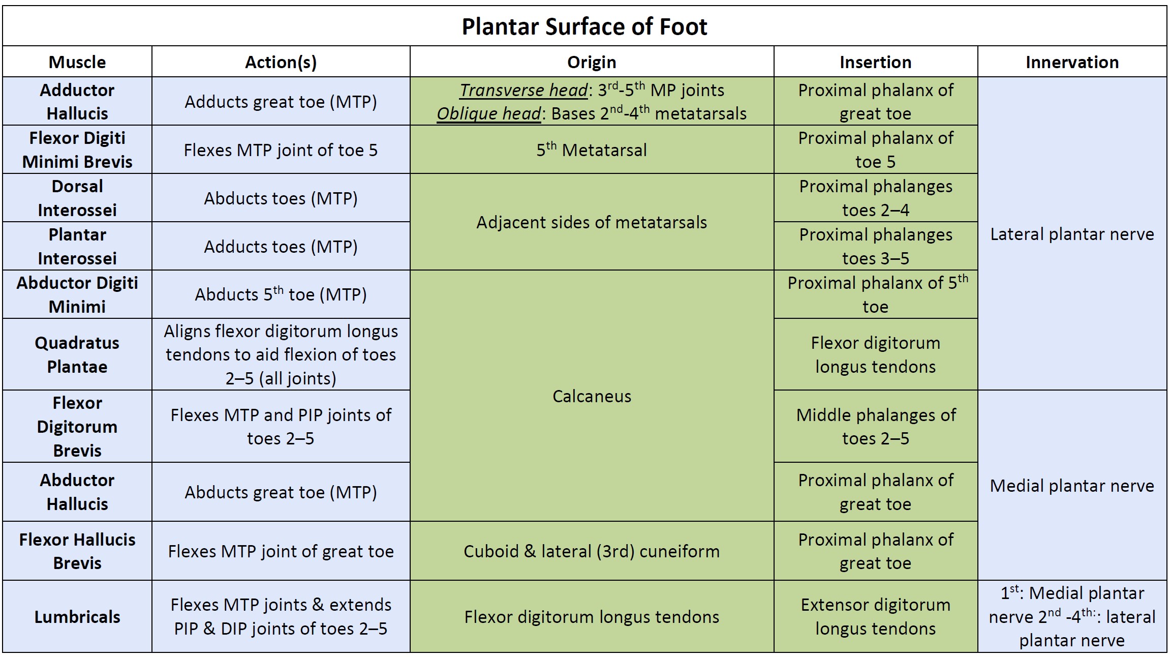
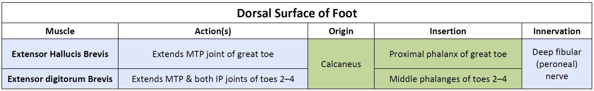
Intrisinc Muscles of the Foot
This content will be covered in lecture.
The intrinsic muscles originate and insert within the foot. These muscles primarily provide support for the foot and its arch, and contribute to movements of the toes.
The dorsal group includes two muscles, the extensor digitorum brevis and extensor hallucis brevis (not labeled on the image below).
The plantar group consists of ten muscles in four layers, and these layers can be used to help you organize the muscles as you study. However, we will not ask you to indicate which layer each muscle is located in.