
Module 31: Upper Extremity III – Elbow and Forearm
Learning Objectives:
By the end of this class, students will be able to:
- List the bones of the arm and elbow joint.
- Identify the bony and ligamentous structures of the elbow joint.
- Establish relationships between bony landmarks and attachments of muscles/tendons or neurovascular structures in the upper extremities.
- Describe the elbow joint and infer how the muscle actions will create movement about the joint.
- Review the innervation of muscles of the arm
Terms to Know
|
Bones
|
Muscles of the Arm
Muscles of the Forearm and Wrist
|
*NOTE – The table below contains the same information from M30. However, now the only bones presented are the humerus, radius, and ulna.
Bones and Bony Landmarks of the Arm and Forearm
*NOTE – The table below contains the same information from M30. However, now the only bones presented are the humerus, radius, and ulna.
| Bone | Bony Landmark | Associated Structures |
| Humerus | Head | Articulates with the glenoid fossa of the scapula |
| Anatomical neck | Just distal to the head of the humerus | |
| Surgical neck | Common site for fractures of the humerus | |
| Deltoid tuberosity | Attachment for the deltoid muscle | |
| Lesser tubercle | Attachment for the subscapularis muscle (and teres major) | |
| Greater tubercle | Attachment for the supraspinatus, infraspinatus, and teres minor | |
| Intertubercular sulcus (groove) | Passageway for the tendon of the long head of the biceps; attachment for the latissimus dorsi and teres major muscles | |
| Radial groove | Radial nerve travels here on its course around the posterior humerus | |
| Medial epicondyle | Attachment site for muscles of the anterior forearm (flexors and pronator teres) | |
| Lateral epicondyle | Attachment site for muscles of the posterior forearm (extensors and supinator) | |
| Lateral supracondylar ridge | Attachment site for some posterior forearm muscles | |
| Medial supracondylar ridge | Attachment site for some anterior forearm muscles | |
| Capitulum | Articulates with the head of the radius | |
| Trochlea | Articulates with the ulna | |
| Radial fossa | Space for radius during elbow flexion | |
| Coronoid fossa | Space for the coronoid process of the ulna during elbow flexion | |
| Olecranon fossa | Space for the olecranon process of the ulna during elbow extension | |
| Ulna | Trochlear notch | Articulates with the trochlea of the humerus |
| Olecranon process | Attachment for the triceps brachii | |
| Coronoid process | Attachment for ligaments of the elbow | |
| Radial notch | Proximal articulation with the head of the radius | |
| Head | Articulates distally with the radius and with carpal bones | |
| Styloid process | Attachment for a ligament of the wrist | |
| Radius | Head | Articulates with the ulna and humerus |
| Radial tuberosity | Attachment for the biceps brachii tendon | |
| Styloid process | Attachment site for the brachioradialis muscle | |
| Ulnar notch | Distal articulation with the ulna |
Joints of the elbow and forearm
This content will be covered in the assignment and reviewed in lecture.
| Joints of the Elbow and Forearm | |||
| Joint | Bones Articulating | Classification | Motions Available |
| Humeroulnar (Elbow) Joint | Trochlea of the humerus and the trochlear notch of the ulna | Hinge Joint | Flexion/Extension |
| Humeroradial (Elbow) Joint | Capitulum of the humerus and the radial head | Hinge Joint | Flexion/Extension |
| Proximal Radioulnar Joint | Head of the radius and the radial notch of the ulna | Pivot Joint | Rotation (pronation/supination) |
| Distal Radioulnar Joint | Head of the ulna and the ulnar notch of the radius | Pivot Joint | Rotation (pronation/supination) |
Elbow Joint
This content will be covered in lecture.
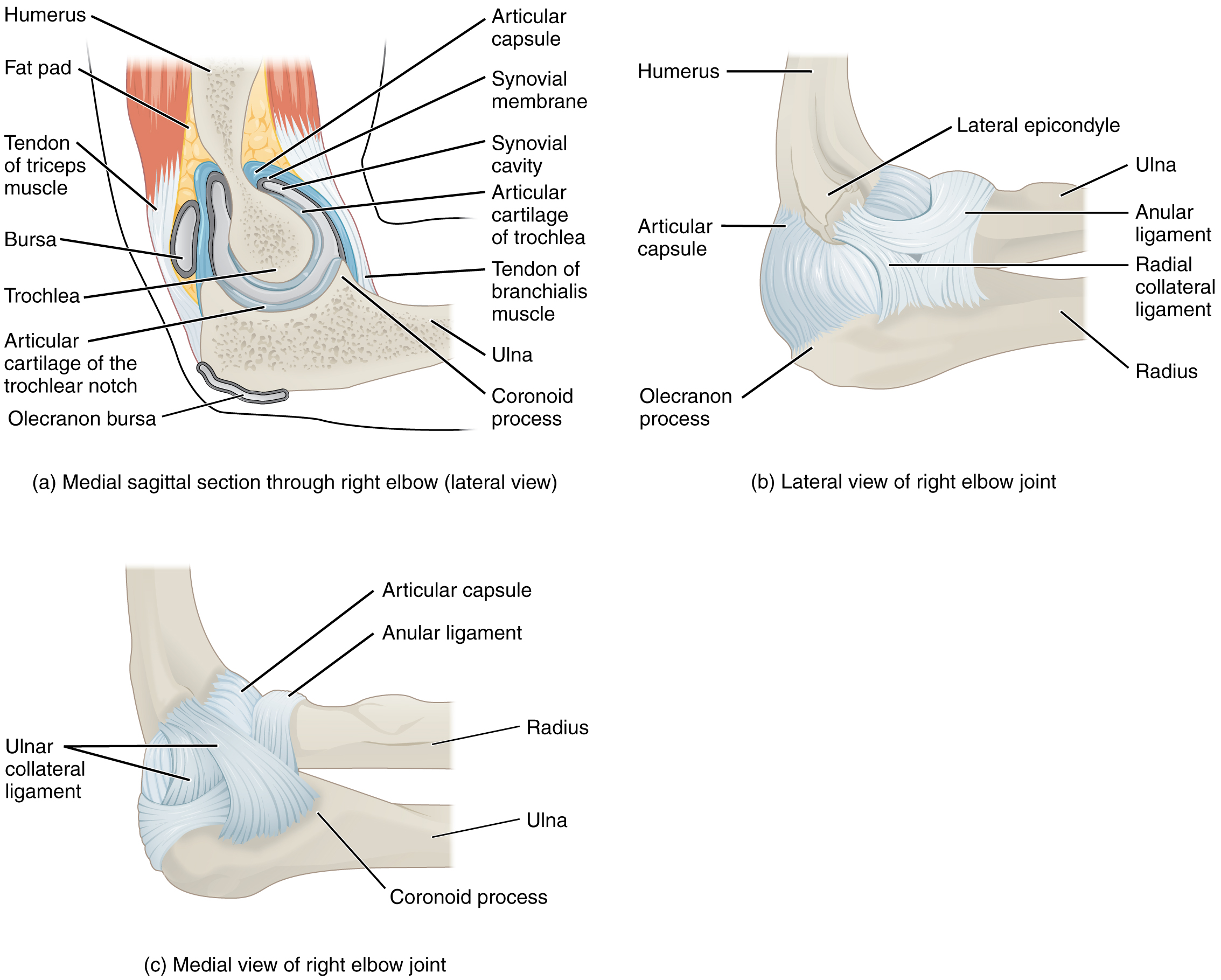
The elbow joint is a uniaxial hinge joint formed by the humeroulnar joint, the articulation between the trochlea of the humerus and the trochlear notch of the ulna. Also associated with the elbow are the humeroradial joint and the proximal radioulnar joint. All three of these joints are enclosed within a single articular capsule.
The articular capsule of the elbow is thin on its anterior and posterior aspects, but is thickened along its outside margins by strong intrinsic ligaments. These ligaments prevent side-to-side movements and hyperextension. On the medial side is the triangular ulnar collateral ligament. This arises from the medial epicondyle of the humerus and attaches to the medial side of the proximal ulna. The strongest part of this ligament is the anterior portion, which resists hyperextension of the elbow.
Ulnar Collateral Ligament – Fun Fact (-> you will not be assessed on this content)
The ulnar collateral ligament may be injured by frequent, forceful extensions of the forearm, as is seen in baseball pitchers. Reconstructive surgical repair of this ligament is referred to as Tommy John surgery, named for the former major league pitcher who was the first person to have this treatment.
The lateral side of the elbow is supported by the radial collateral ligament. This arises from the lateral epicondyle of the humerus and then blends into the lateral side of the annular ligament. The annular ligament encircles the head of the radius. This ligament supports the head of the radius as it articulates with the radial notch of the ulna at the proximal radioulnar joint. This is a pivot joint that allows for the rotation of the radius during supination and pronation of the forearm.
Elbow Joint
Radioulnar Joints
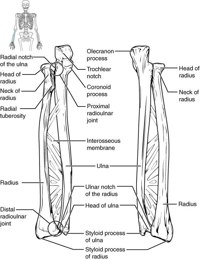
There are two articulations between the radius and ulna called the proximal radioulnar joint and the distal radioulnar joint. The proximal radioulnar joint is the articulation between the head of the radius and the radial notch of the ulna. The distal radioulnar joint is the articulation between the head of the ulna and the ulnar notch of the radius. A dense connective tissue that unites the ulna and radius is called the interosseous membrane. The radioulnar joints are pivot joints, allowing for the forearm motions of pronation and supination.
Ulna and Radius
Muscles That Move the Forearm
This content will be covered in the assignment and reviewed in lecture.
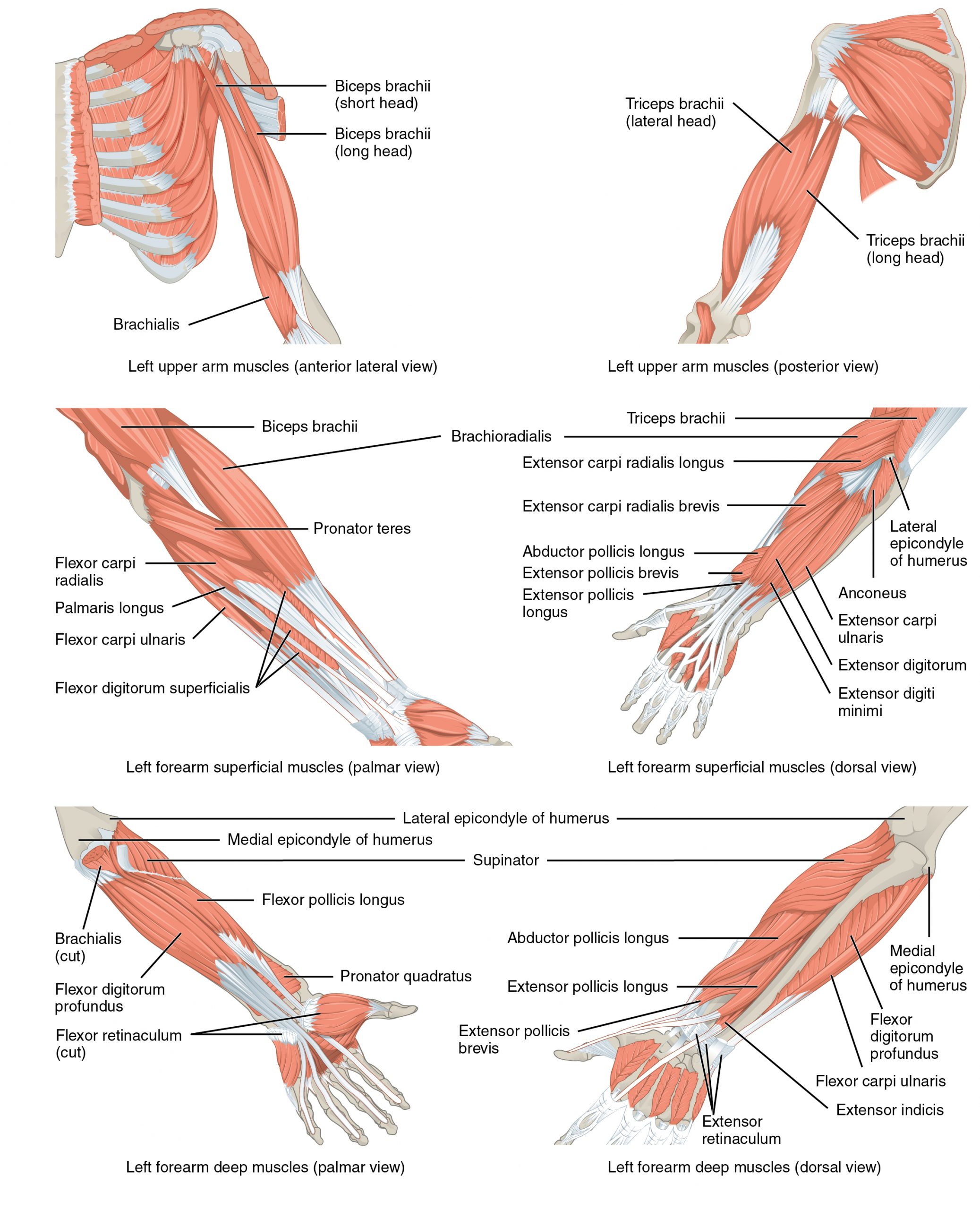
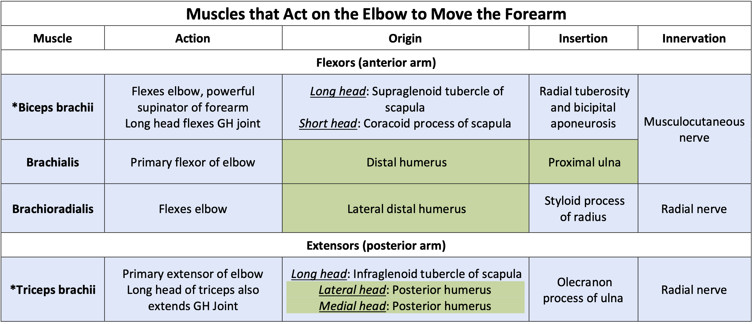
The forearm, made of the radius and ulna bones, has four main types of action: flexion and extension occur at the elbow joint, whereas pronation and supination occur between the radius and ulna. The elbow flexors include the biceps brachii, brachialis, and brachioradialis. The two-headed biceps brachii crosses the shoulder and elbow joints, causing shoulder and elbow flexion. Also, the biceps brachii is the primary supinator of the forearm. The primary elbow extensor is the triceps brachii. The pronators are the pronator teres and the pronator quadratus, and the supinator is the only one that turns the palm anteriorly. When the palm faces anteriorly, it is supinated. When the palm faces posteriorly, it is pronated.
Muscles That Move the Forearm
Use the image/table below to understand the actions, origins, insertions, and innervation of the muscles that move the elbow and forearm.
*Note: Any box shaded in blue on the muscle chart requires you to know all the information (e.g., action, origin, insertion, and innervation) provided. Areas shaded in green require muscle identification and a general sense of the location of the origin or insertion. (e.g., knowing that the rhomboids originate on the vertebrae, as opposed to the specific numbered vertebrae).
Muscles That Move the Wrist, Hand, and Fingers
This content will be covered in the assignment and reviewed in lecture.
Wrist, hand, and finger movements are facilitated by two groups of muscles. The forearm is the origin of the extrinsic muscles of the hand. The palm is the origin of the intrinsic muscles of the hand.
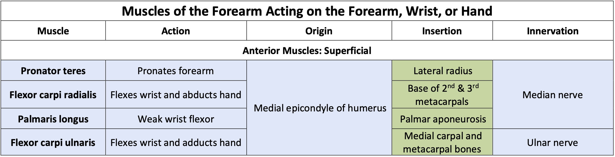
Muscles of the Forearm That Move the Wrists, Hands, and Fingers
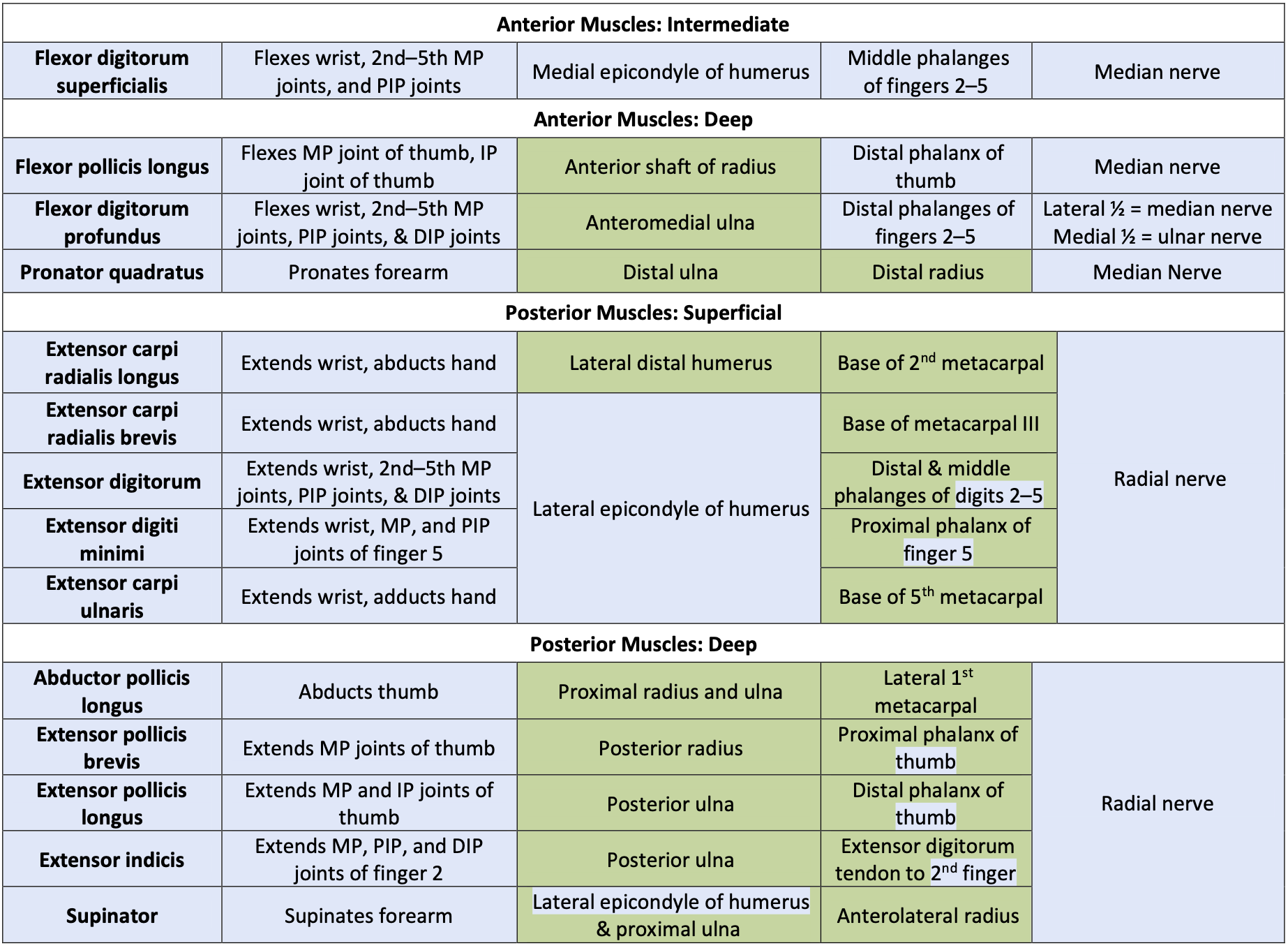
The muscles in the anterior compartments of the forearm originate on the humerus and insert onto different parts of the forearm and hand. From lateral to medial, the superficial anterior compartment of the forearm includes the pronator teres, flexor carpi radialis, palmaris longus, and flexor carpi ulnaris. The intermediate anterior compartment is comprised of only the flexor digitorum superficialis, a wrist, hand, and finger flexor. The deep anterior compartment is comprised of three muscles, the flexor pollicis longus, flexor digitorum profundus, and pronator quadratus.
- Every muscle of the anterior forearm is innervated by the Median Nerve
- **EXCEPT** – flexor carpi ulnaris and the medial 1/2 of flexor digitorum profundus – these are innervated by the Ulnar Nerve
The muscles in the superficial posterior compartment of the forearm originate on the humerus. These are the extensor carpi radialis longus, extensor carpi radialis brevis, extensor digitorum, extensor digiti minimi, and the extensor carpi ulnaris. The muscles of the deep posterior compartment of the forearm originate on the radius and ulna. These include the abductor pollicis longus, extensor pollicis brevis, extensor pollicis longus, extensor indicis, and the supinator.
- Every muscle of the posterior forearm is innervated by the Radial Nerve
- *Note* – The radial nerve also innervates the brachioradialis and triceps brachii
The tendons of the forearm muscles attach to the wrist or extend into the hand. Fibrous bands called retinacula sheath the tendons at the wrist. The flexor retinaculum extends over the palmar surface of the hand while the extensor retinaculum extends over the dorsal surface of the hand.
