
Lab 6: BACKGROUND
Site-Directed Mutagenesis
By completing labs 2, 4, and 5, we have now cloned the wild type HCAII gene into the pETblue-2 vector. In order to study our mutations of interest, we next need to take that wild type clone and introduce mutations. In 551 we use accomplish this using a protocol called site-directed mutagenesis.
Please review the site-directed mutagenesis section of the PCR & Cloning lectures before lab.
Conservation Analysis: Sequence Alignment
A sequence alignment is a computational tool that arranges sequences to identify regions of similar or even matching sequence. These alignments can be used for a number of purposes, such as identifying evolutionary relationships between different protein sequences. In the case of a mutational analysis, such as we are doing in 551, it can be useful to do a sequence alignment of protein sequences from across different species. This can provide information on whether a given amino acid residue is evolutionarily conserved – i.e., whether the same amino acid is present at the same position across different versions of the protein. If an amino acid is evolutionarily conserved, that suggests that it plays an important role in the protein. If an amino acid is not conserved, it may be less important. Some amino acids are partially conserved, which means that different amino acids are found at that position, but those amino acids all have similar properties – i.e., negative charge.
Below is a sample image of a sequence alignment:
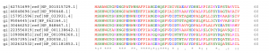
You can see that nine sequences have been aligned, and each amino acid is color-coded according to biochemical property (for example, negatively charged residues are blue). The first amino acid is, of course, methionine, which is completely conserved, as indicated by the “*” at the bottom of the column. The second amino acid is highly conserved, denoted with a “:” because most of the sequences have a serine, but two have an alanine. Resides denoted with a “.” are considered mostly conserved. Jumping forward to the eighth position, you can see that this position is not conserved at all, as sequences may contain a glycine, alanine, serine, or aspartic acid here.
How is this useful to studying mutations? If you were proposing to mutate this protein at the eighth amino acid, you might anticipate this having little effect, since the protein seems to tolerate many different amino acids at this position. By contrast, the third amino acid is a completely conserved histidine. If you were proposing to mutate this histidine, you might be more likely to expect some effect. (Of course, sequence conservation is just one piece of information to take into account when forming a hypothesis. The role of the wild type amino acid in the protein, its proximity to the active site, and the biochemical properties of both the wild type and mutated residues are also essential considerations.)
Making an Informative PyMOL Figure
You already have some experience with making nice PyMOL figures, and in this lab you will be applying those principles to creating images that help an audience understand your mutation.
The video linked below includes a number of tips and tricks for creating useful PyMOL figures:
